Science is beautiful!
In our research we do not only answer important scientific questions, but also generate beautiful images on the tissues and proteins that we work with. We’ve used a selection of them as backgrounds on our website. Have a look below for a brief explanation.


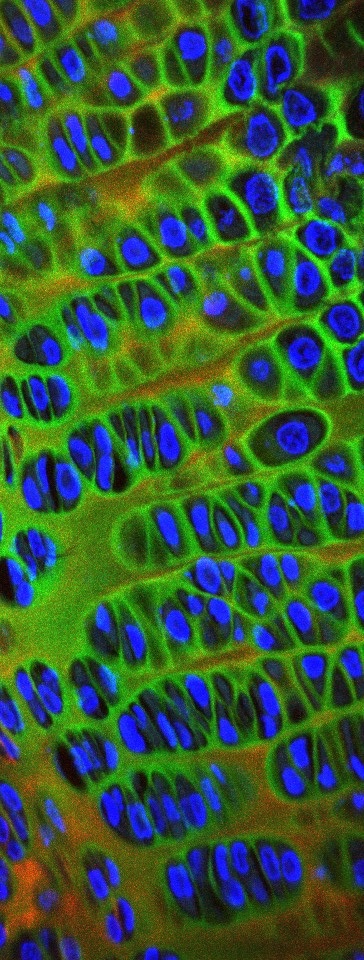
Immunohistochemistry for matrilin 3 (green) and type II collagen (red) in the cartilage growth plate. The nuclei are stained blue (DAPI). Image by Dr Peter Bell.

3D reconstruction of a chondron from mice harbouring V194D matrilin-3 mutation (spot the enlarged ER inside the cells, full of mutant protein!). Image by Dr Matthew Leighton.
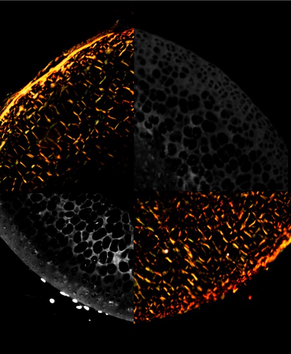
A collage of Picrosirus Red (observed under polarised light, thick collagen bundles in orange, thin in yellow and green) and DAPI (slightly overexposed; grey) staining of decellularised (bottom half) and cell-seeded decellularised (top half) murine femoral heads. Image by Dr Katarzyna Pirog.
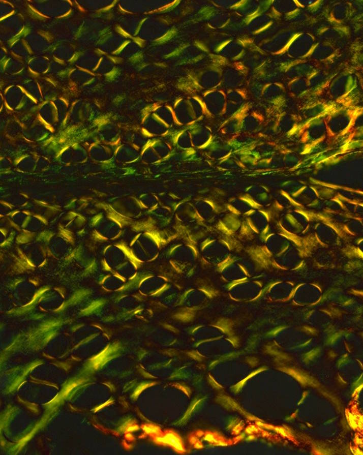
Picrosirus red stained articular cartilage (femur and tibia) observed under polarised light. Thick collagen fibres in red, thin in green. Image by Roufaida Bouchenafa (PhD student).
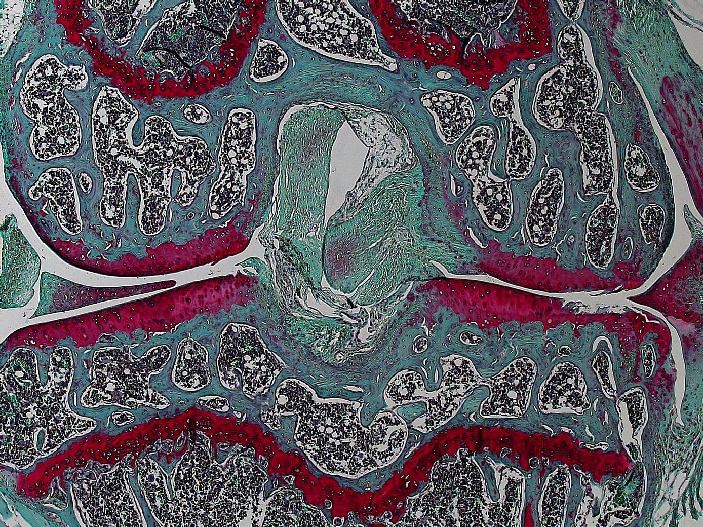
Safranin O staining of mouse tibial joint. Cartilage in red, bone in blue. Image by Dr Katarzyna Pirog.
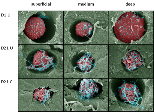
Scanning electron micrographs (SEM) of ATDC5 cells cultured in 2% agarose with (C) or without (UC) compression. Agar coloured in green, cells in red, pericellular matrix in blue. Image by Dr Marc Farcasanu.