Getting into the ‘flow’

So we’ve had our summer holiday fun, now to get back into “the flow” of the lab! 😉 (not that we haven’t been working hard over the summer too!). We have lots planned coming up, so keep an eye out for a range of updates!

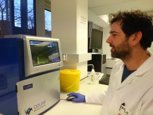
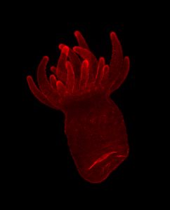
One thing we have been working on is practicing some removal assays! what are they I hear you ask? Simply, we attach some anemones to slides of different surface chemistries/textures, put them in a machine which flows water over them at specified flow rates, and we see how many are stuck on at the end! Here is a little clip of some of our anemones in the flow cell. Keep an eye out for the guy in the middle of the middle slide. You can see it peel off in stages!

Now, why on earth would we need to do this? whats the point? the first aspect is part of seeing how strongly they adhere to surfaces. This will be combined with other measurements in other experiments to give a good idea of just how strong they stick and how good their glue is!

The other component of this particular experiment is to see how their ability to stick to DIFFERENT surfaces changes. Maybe they stick better to one type compared to another? This will contribute to characterizing their adhesion, but also, seeing as we are part of a bio-fouling research group!; we can see which type of surfaces are less likely to be colonized by this species. This is important information when you don’t want sea life growing all over your boat or other underwater structures.

The sweet side of adhesives

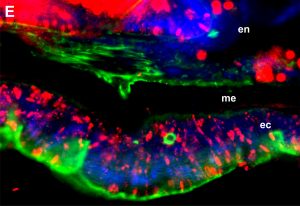
It is well known, natural adhesives contain sugars (glycan) in its composition. These sugars are post-translationally attached to the protein backbone (glycoproteins) and are predicted to play an important role in underwater attachment – this isn’t different in sea anemones. Therefore, understanding these glycoproteins is fundamental to our project. One way to study them is by using a method called immuno-histochemistry, which combines anatomical, immunological, biochemical and microscopy techniques to image cellular components. Lectins are glycan-binding proteins that are highly specific for glycan-conjugates and can be used to target sugar moieties in-situ. By combining immuno-histochemistry and fluorescence-labeled lectins, it is possible to visualize and document the distribution and localization of glycoproteins. We started fiddling with this technique in Aiptasia and the results are AWESOME. We couldn’t help ourselves but share some images from our experiment. 

With this method, we can use different fluorescent and light microscopy channels: A) bright field illumination. en= endoderm, me= mesoglea, ec: ectoderm. The ectoderm in the image depicts basal disc cells which participate in the attachment of the sea anemones to the surfaces, therefore, these cells secrete the glue molecules we are investigating; B) lectin staining using Texas red as the secondary antibody. This is the actual distribution of glycoconjugates in these tissues; C) actin filaments labeled with Phalloidin; D) DNA staining using DAPI.

When we overlay all the different channels we get this pleasing to look at image (E). We are still exploring the full potential of lectin staining and will keep experimenting in this area.


ENBA (European Network of Bioadhesion) march meeting

Image

It has been a great experience attending the COST Action ENBA (European Network of Bioadhesion) meeting in Barcelona March 5-6th. Two members of the (A)nemone team attended the meeting to discuss on the bio-inspired adhesives. Nick as a core member and a ‘working group 1’ leader attended both days, while Marcelo joined socially for the dinner and formally for the second day (Yes, Jess stayed in Newcastle nursing our anemones and making sure all was running well). The first day was for core members only to undertake executive decisions. On the second day, we enjoyed presentations from ENBA members and networking with international colleagues. The most enriching experience of the meeting was with no doubt to meet with old colleagues and to make new ones. Great opportunities have arisen from the discussions to create a Biomolecular focused working group and an Early Career Group within ENBA’s main activities. Thank you very much to all that made the meeting possible. We are looking forward to meeting with you at the next meeting, wherever it is!

For more information about ENBA, see their website: http://www.enba4.eu/

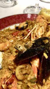
Barnacles attached to mussels and all found in a paella during dinner. That’s a great example of bioadhesion!

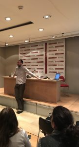
Nick as a session chair during presentations.

Happy new year and project update!

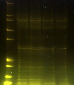
Busy start to 2018 already, and we have kick started with successful protein extractions of our anemones! Next steps will include a few more extractions from different parts of the animal as well as the footprint (the glue left behind), in order to seek out any specific proteins involved in adhesion. We will also be doing Mass spectrometry! Mass spectrometry will give us vital information to identify the extracted proteins. We are looking forward to doing some collision-induced dissociation of these peptides. Freaaaky!

Happy New Year from Newcastle!


Big day for Team Anemone on Friday December 15th…

…yes, the launch of our long-awaited blog!  Just in time for Christmas.

Okay, we officially kicked-off our first big project in May (Leverhulme Research Project Grant) so it’s taken us some time, but a lot has been happening!

After Marcelo arrived in May 2017 we established cultures of everybody’s favourite anemone, Aiptasia, with lots of help from the Guse Lab in Heidelberg – thank you Annika et al. 😉  Jess followed in September and the A-Team is now up to full strength.  We have an exciting 4 years ahead – so many opportunities, so little time.  It has taken a lot of hard work by Marcelo and myself to get us to this point…  I started investigating adhesion of sea anemones during a 2012 Visiting Professorship to St FX university in Nova Scotia.  Marcelo was simultaneously embarking on a Marie Curie Fellowship at the University of Innsbruck, studying adhesion of Hydra.  Our paths crossed at a COST Action bioadhesion meeting in Istanbul in 2014 and the rest is history!  Jess is new to all of this adhesion stuff, but she is already getting ‘stuck in’ (ha ha….).

Stay tuned for updates and exciting news from the worlds of anemones and bioadhesion,

Nick Aldred, Marcelo Rodriguez and Jess Clarke