Getting into the ‘flow’

So we’ve had our summer holiday fun, now to get back into “the flow” of the lab! 😉 (not that we haven’t been working hard over the summer too!). We have lots planned coming up, so keep an eye out for a range of updates!

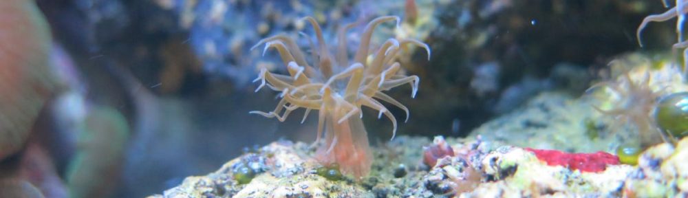
One thing we have been working on is practicing some removal assays! what are they I hear you ask? Simply, we attach some anemones to slides of different surface chemistries/textures, put them in a machine which flows water over them at specified flow rates, and we see how many are stuck on at the end! Here is a little clip of some of our anemones in the flow cell. Keep an eye out for the guy in the middle of the middle slide. You can see it peel off in stages!

Now, why on earth would we need to do this? whats the point? the first aspect is part of seeing how strongly they adhere to surfaces. This will be combined with other measurements in other experiments to give a good idea of just how strong they stick and how good their glue is!

The other component of this particular experiment is to see how their ability to stick to DIFFERENT surfaces changes. Maybe they stick better to one type compared to another? This will contribute to characterizing their adhesion, but also, seeing as we are part of a bio-fouling research group!; we can see which type of surfaces are less likely to be colonized by this species. This is important information when you don’t want sea life growing all over your boat or other underwater structures.

The sweet side of adhesives

It is well known, natural adhesives contain sugars (glycan) in its composition. These sugars are post-translationally attached to the protein backbone (glycoproteins) and are predicted to play an important role in underwater attachment – this isn’t different in sea anemones. Therefore, understanding these glycoproteins is fundamental to our project. One way to study them is by using a method called immuno-histochemistry, which combines anatomical, immunological, biochemical and microscopy techniques to image cellular components. Lectins are glycan-binding proteins that are highly specific for glycan-conjugates and can be used to target sugar moieties in-situ. By combining immuno-histochemistry and fluorescence-labeled lectins, it is possible to visualize and document the distribution and localization of glycoproteins. We started fiddling with this technique in Aiptasia and the results are AWESOME. We couldn’t help ourselves but share some images from our experiment. 

With this method, we can use different fluorescent and light microscopy channels: A) bright field illumination. en= endoderm, me= mesoglea, ec: ectoderm. The ectoderm in the image depicts basal disc cells which participate in the attachment of the sea anemones to the surfaces, therefore, these cells secrete the glue molecules we are investigating; B) lectin staining using Texas red as the secondary antibody. This is the actual distribution of glycoconjugates in these tissues; C) actin filaments labeled with Phalloidin; D) DNA staining using DAPI.

When we overlay all the different channels we get this pleasing to look at image (E). We are still exploring the full potential of lectin staining and will keep experimenting in this area.