It is well known, natural adhesives contain sugars (glycan) in its composition. These sugars are post-translationally attached to the protein backbone (glycoproteins) and are predicted to play an important role in underwater attachment – this isn’t different in sea anemones. Therefore, understanding these glycoproteins is fundamental to our project. One way to study them is by using a method called immuno-histochemistry, which combines anatomical, immunological, biochemical and microscopy techniques to image cellular components. Lectins are glycan-binding proteins that are highly specific for glycan-conjugates and can be used to target sugar moieties in-situ. By combining immuno-histochemistry and fluorescence-labeled lectins, it is possible to visualize and document the distribution and localization of glycoproteins. We started fiddling with this technique in Aiptasia and the results are AWESOME. We couldn’t help ourselves but share some images from our experiment. 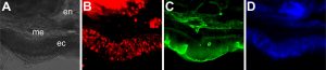
With this method, we can use different fluorescent and light microscopy channels: A) bright field illumination. en= endoderm, me= mesoglea, ec: ectoderm. The ectoderm in the image depicts basal disc cells which participate in the attachment of the sea anemones to the surfaces, therefore, these cells secrete the glue molecules we are investigating; B) lectin staining using Texas red as the secondary antibody. This is the actual distribution of glycoconjugates in these tissues; C) actin filaments labeled with Phalloidin; D) DNA staining using DAPI.
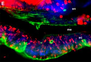
When we overlay all the different channels we get this pleasing to look at image (E). We are still exploring the full potential of lectin staining and will keep experimenting in this area.