By Erin Cocks

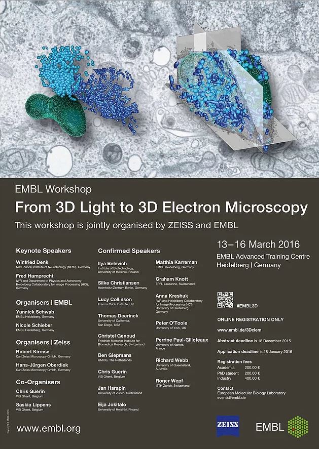
I was recently given the opportunity to attend a workshop on 3D Light and Electron Microscopy and the Gatan 3View Users conference at the European Molecular Biology Laboratory in Heidelberg, Germany. Being my first experience at an academic conference I was excited to hear from those who have helped develop the electron microscopy discipline. From those who pioneered the first Serial-Block Face Scanning Electron Microscope, SBFSEM, to the new and upcoming developments or research being done across the globe. The technique itself allows for serial sectioning and imaging of a block of tissue, which can then be annotated or segmented and 3D reconstructions made of the tissue, like the example below.

An EM orthoslice of Guinea Pig Foetal Skeletal Muscle and the 3D reconstruction of the stack of images (Scale bar is 1µm), taken from my own research. The light blue is mitochondria, green the cell boundary, the pink is chromatin and the dark blue the nucleolus in the nucleus, which has been made transparent.
The topics discussed ranged from neural tissue to plant tissue 3D reconstruction as well as new ways to optimise the processing techniques for specific tissue types. One of the advances in neuronal tissue reconstructing is the ability to now do whole brain staining (Mikula and Denk, 2015). This means that large volumes of tissue can be reconstructed all from the same brain, leading to the reconstruction of an entire mouse brain. This is all part of the ongoing Connectomics project http://www.openconnectomeproject.org/. With these advancements in the technique we will only learn more and more about the brain and many other tissues.
A lot was learnt at the conferences, too much to put in a single blog post, and I am already looking forward to the next one in 2 years time and the other conferences and courses in between.
References
Mikula, S., and Denk, W. (2015). High-resolution whole-brain staining for electron microscopic circuit reconstruction. Nat. Methods 12, 541–546