Category Archives: Publications
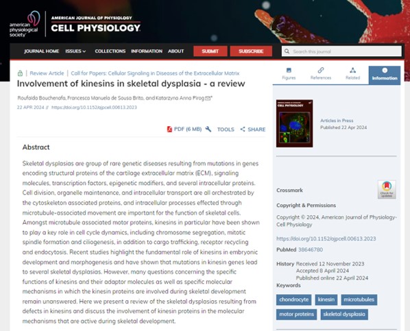
Our review on the role of kinesins in skeletal disease is out
Well done Roufaida and Fran for writing a very comprehensive review of kinesin involvement in skeletal disease. It highlights several novel roles for kinesins in skeletal development and provides great background for our current work.
Full text available here: https://journals.physiology.org/doi/abs/10.1152/ajpcell.00613.2023
mir-324 and hippocampal exitability – a new paper from the Young (SRG) lab
Our new paper is out!
Our new paper, showing an isomiR of miR-140-3p is the most functional in cartilage, is out in RNA
Our new review is now available
For the full paper, please click here: https://doi.org/10.12688/f1000research.22275.1
Enjoy!
Our new paper is out in print!
Well done Ella, great work, and opens many interesting research avenues in the future
A new pre-print from the Skeletal Research Group in Newcastle
A new pre-print from our collaboration with Prof David Young at Newcastle. We helped with the animal work on this project. Click on the link below and tell us what you think!
microRNA-seq of cartilage reveals an over-abundance of miR-140-3p which contains functional isomiRs
Our paper was accepted in PLoS Genetics!
ARMET/MANF paper accepted for publication in Cell Stress and Chaperones
Mesencephalic astrocyte-derived neurotrophic factor is an important factor in chondrocyte ER homeostasis.
Bell PA, Dennis EP, Hartley CL, Jackson RM, Porter A, Boot-Handford RP, Pirog KA, Briggs MD.
Mesencephalic astrocyte-derived neurotrophic factor (MANF) is an endoplasmic reticulum (ER) resident protein that can be secreted due to an imperfect KDEL motif. MANF plays a cytoprotective role in several soft tissues and is upregulated in conditions resulting from intracellular retention of mutant protein, including two skeletal diseases, metaphyseal chondrodysplasia type Schmid (MCDS) and multiple epiphyseal dysplasia (MED).
The role of MANF in skeletal tissue homeostasis is currently unknown. Interestingly, cartilage-specific deletion of Manf in a mouse model of MED resulted in increased disease severity, suggesting its upregulation may be chondroprotective. Treatment of MED chondrocytes with exogenous MANF led to a decrease in the cellular levels of BiP (GRP78), confirming MANF’s potential to modulate ER stress responses. However, it did not alleviate the intracellular retention of mutant matrilin-3, suggesting that it is the intracellular MANF that is of importance in the pathobiology of skeletal dysplasias.
The Col2Cre-driven deletion of Manf from mouse cartilage resulted in a chondrodysplasia-like phenotype. Interestingly, ablation of MANF in cartilage did not have extracellular consequences, but led to an upregulation of several ER-resident chaperones including BiP. This apparent induction of ER-stress in turn led to dysregulated chondrocyte apoptosis and decreased proliferation, resulting in reduced long bone growth.
We have previously shown that ER stress is an underlying disease mechanism for several skeletal dysplasias. The cartilage-specific deletion of Manf described in this study phenocopies our previously published chondrodysplasia models, further confirming that ER-stress itself is sufficient to disrupt skeletal growth and thus represents a potential therapeutic target.
First paper of 2018 published!
Calcium activated nucleotidase 1 (CANT1) is critical for glycosaminoglycan biosynthesis in cartilage and endochondral ossification.
Desbuquois dysplasia type 1 (DBQD1) is a chondrodysplasia caused by mutations in CANT1 gene encoding an ER/Golgi calcium activated nucleotidase 1 that hydrolyses UDP. Here, using Cant1 knock-in and knock-out mice recapitulating DBQD1 phenotype, we report that CANT1 plays a crucial role in cartilage proteoglycan synthesis and in endochondral ossification. Specifically, the glycosaminoglycan synthesis was decreased in chondrocytes from Cant1 knock-out mice and their hydrodynamic size was reduced, whilst the sulfation was increased and the overall proteoglycan secretion was delayed. Interestingly, knock-out chondrocytes had dilated ER cisternae suggesting delayed protein secretion and cellular stress; however, no canonical ER stress response was detected using microarray analysis, Xbp1 splicing and protein levels of BiP and ATF4. The observed proteoglycan defects caused deregulated chondrocyte proliferation and maturation in the growth plate resulting in the reduced skeletal growth. In conclusion, the pathogenic mechanism of DBQD1 comprises deregulated chondrocyte performance due to defective intracellular proteoglycan synthesis and altered proteoglycan properties in the extracellular matrix.