PolyU collaborators visit Newcastle to conduct joint microneedle study

2023 seemingly left in a haste. Stepping into 2024, we welcomed our collaborators from The Hong Kong Polytechnic University (PolyU) to Newcastle, to conduct a joint study on microneedle formulation for drug delivery and diagnostics. Merab Naveed, Hubert Chan and Dr Thomas Lee from PolyU’s Biomedical Engineering Department spent nearly two weeks with us, running experiments and exchanging ideas with us. Newcastle University students, Begho Obale and Jakub Masloch, who completed their MPharm research projects with us, also lent their expertise to this joint study. Among other things, Begho made a dancing microneedle mould – the first ever reported. It was a most wonderful way to start the new year.

So how did our guests find it? I know Hubert enjoyed the unique learning experience – his words, not mine. I’m really pleased that we’re able to organise this research exchange programme. Thanks also go to Dr Wing Man Lau and Dr Hin Chung Lau of PolyU, the other two academic advisors on the project, for making this happen.

Perspective: The diagnostic potential of microneedles in infectious diseases

The recent COVID-19 pandemic brought the healthcare systems in many countries to their knees. One of the key problems that became evident early on was the lack of diagnostic tools that were rapid and readily accessible to the public.

Diagnostics is what we do as a research team. Naturally, we contemplated what we could do in a situation like this. The result is a perspective article published in Precision Nanomedicine, the official journal of the European Foundation for Nanomedicine.

The conclusion? Microneedle systems excel in many ways as a diagnostic tool. Various microneedle platforms have demonstrated advantages of portability, self-administrability, affordability and scalability over other diagnostic platforms. The potential is huge but the technology is still in its infancy. We need more research to turn that potential into tangible clinical benefits, but we can’t do it alone. Cross-disciplinary collaboration and stakeholder support will be needed to drive this effort forward.

The article is published under the Creative Commons BY-NC-SA 4.0 licence, so please feel free to distribute widely, adapt and reuse for non-commercial purposes, and share any derived work, citing:

Dixon RV, Lau WM, Moghimi SM, Ng KW (2020) The diagnostic potential of microneedles in infectious diseases. Precision Nanomedicine 3(4):629–640 . https://doi.org/10.33218/001c.13658

A microneedle array biosensor that can detect skin burns

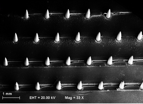
SEM image of microneedle array biosensor

We have developed a microneedle array biosensor that can detect skin burns. To make the biosensor, we devised a nanocomposite material from carbon nanotubes and a biocompatible polymer, poly(lactic acid). We then shaped the microneedle array from the nanocomposite material. The microneedles that formed were about 870 μm long, 250 μm wide, and electroactive. This meant that the microneedle array could be inserted into  the skin to detect certain analytes by electrochemistry. We verified this by using the microneedle array to detect vitamin C in solution. Interestingly, when we tested the microneedle array biosensor on skin burns and normal (non-burnt) skin, the skin specimens showed different electrochemical responses. This gives us the technological basis for a minimally invasive biosensing approach to detecting skin burns.

For more information, please see:

Skaria E, et al. (2019) Poly(lactic) acid/carbon nanotube composite microneedle arrays for dermal biosensing. Analytical Chemistry (epub ahead of print). doi: 10.1021/acs.analchem.8b04980