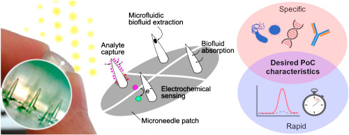
Perspective: The diagnostic potential of microneedles in infectious diseases

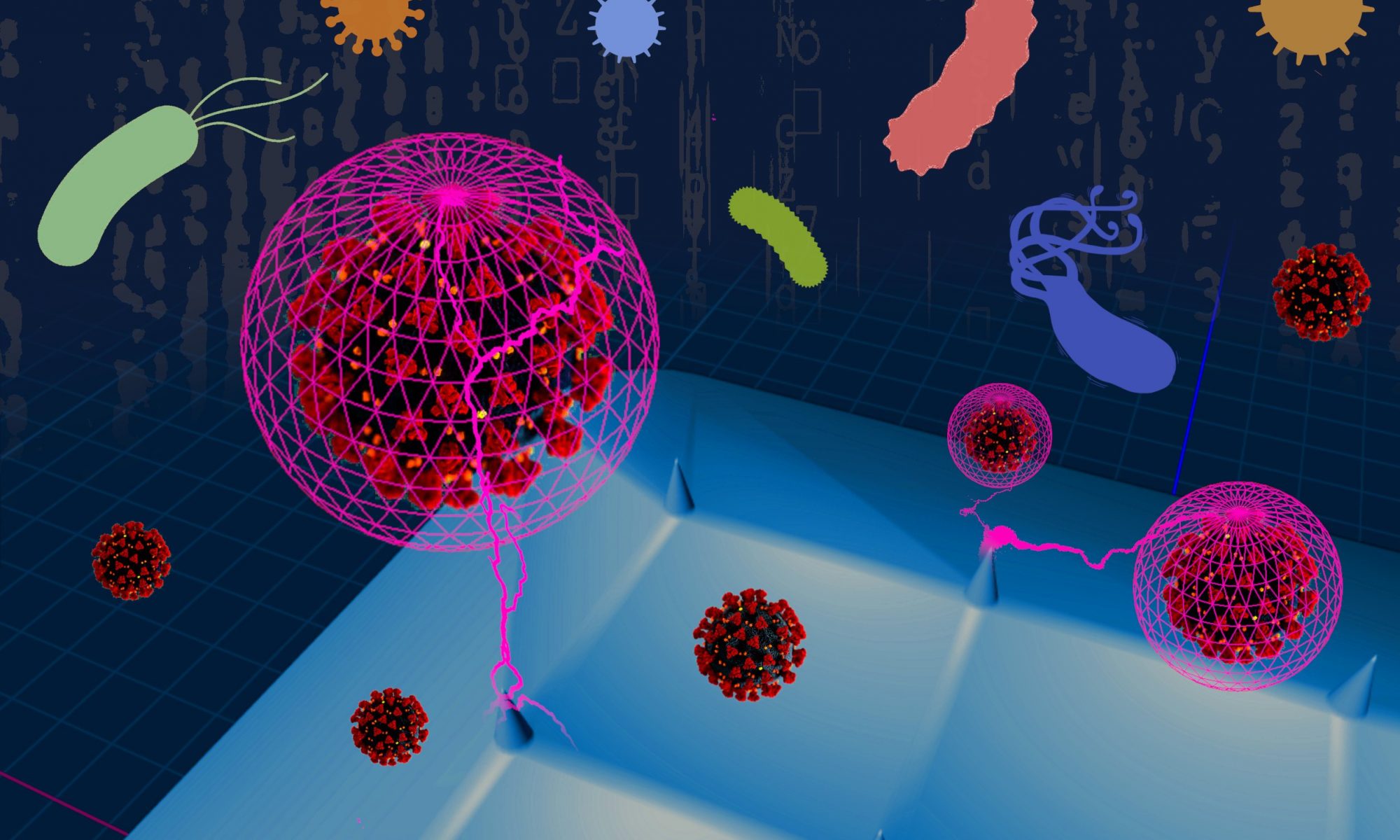
The recent COVID-19 pandemic brought the healthcare systems in many countries to their knees. One of the key problems that became evident early on was the lack of diagnostic tools that were rapid and readily accessible to the public.

Diagnostics is what we do as a research team. Naturally, we contemplated what we could do in a situation like this. The result is a perspective article published in Precision Nanomedicine, the official journal of the European Foundation for Nanomedicine.

The conclusion? Microneedle systems excel in many ways as a diagnostic tool. Various microneedle platforms have demonstrated advantages of portability, self-administrability, affordability and scalability over other diagnostic platforms. The potential is huge but the technology is still in its infancy. We need more research to turn that potential into tangible clinical benefits, but we can’t do it alone. Cross-disciplinary collaboration and stakeholder support will be needed to drive this effort forward.

The article is published under the Creative Commons BY-NC-SA 4.0 licence, so please feel free to distribute widely, adapt and reuse for non-commercial purposes, and share any derived work, citing:

Dixon RV, Lau WM, Moghimi SM, Ng KW (2020) The diagnostic potential of microneedles in infectious diseases. Precision Nanomedicine 3(4):629–640 . https://doi.org/10.33218/001c.13658

Lab hiatus

In the face of the COVID-19 pandemic, we have temporarily shut down all lab experiments until further notice. All team members are following government advice to work from home. We will be monitoring the situation closely and hope to be back in operation as soon as the situation improves. In the meantime, we will be working on those overdue papers…

Skin bank

This week, we achieved a major milestone in our research capabilties. We established a skin bank to ensure a steady supply of skin samples for our research. At present, the skin bank contains porcine and murine skin samples. We may expand this to include other skin samples in the future.

The team was understandably thrilled when we received our first skin donations to the skin bank. Hassan, Rach and Trevor all had a go at processing the skin samples for storage. These skin samples will be put to good use in the coming weeks and months.

Here’s Rach, the self-proclaimed pig barber at work:

Welcoming Hassan

Just as we were about to wind down our lab operations for Christmas, we welcomed postdoctoral research associate, Dr Hassan Elsana, into our team this week. Hassan will be working on an EPSRC-funded project researching microneedle-mediated drug delivery in the skin.

This project is a collaboration with Dr Wing Man Lau (School of Pharmacy) and Dr Katarina Novakovic (School of Engineering). We have high hopes for this project.

Exciting times ahead, and I don’t just mean Christmas!

Welcome, team!

This week, I welcome 4 new team members into the team:

Cerys Evans is an MPharm student who will be investigating microneedle coating strategies for dermal drug delivery over the next 10 weeks.

Rachael Dixon joins us to develop a novel skin health monitor, in a joint effort between the School of Pharmacy and the Institute of Cellular Medicine. Professor Syed Moein Moghimi, Professor Mark Birch-Machin and Dr Philip Manning are co-supervisors.

Grace Young starts her PhD research formulating an antimicrobial delivery strategy to combat biofilms. The project, led by Dr Wing Man Lau and co-supervised by Dr Nick Jakubovics and myself, is a collaboration between the School of Pharmacy and the School of Dental Sciences.

Georgia Peavoy joins us to develop a novel diagnostic device for detecting nerve damage in diabetic patients. This is a collaborative project with Dr Roger Whittaker (lead supervisor, Institute of Neuroscience) and Professor Anthony O’Neil (School of Engineering).

I hope they enjoy their time with us and have a productive research project in the months and years ahead.

4DLab at PharmSci 2019

If you have been keeping up with my tweets, you may have noticed that we were at the 10th APS International PharmSci conference last week in Greenwich, UK.

We presented our collaborative work with Unversity of Surrey’s BioProChem group on our recently published work on microneedle immunodiagnostics, as well as our investigation into the effects of oxygen and pressure on wound healing with Dr Wing Man Lau, Professor Adrian Williams and Dr Kawa Ahmad.

Our MPharm alumni have much to be proud of too. Joanna’s work on microneedle hydrogel coating was presented as a poster, while Arun’s work on a dual-analysis immunodiagnostic microneedle platform for skin cancer detection was presented as part of a podium presentation.

I spoke on day 2 of the conference.

All in all it was a fun and extremely fruitful conference. We look forward to the 11th iteration of the conference in Belfast next year!

Collaborative paper on 3D cell culture for evaluating biomarker-capturing microneedle devices

Update: Author manuscript available

One of the challenges in developing microneedle devices to capture disease biomarkers from the skin is the lack of suitable skin specimens in which to test the devices. Donor skin specimens that carry the specific target diseases simply do not come by easily.

I first discussed this challenge with Eirini Velliou, Tao Chen and Guoping Lian in April 2016. We decided to tackle it by growing our own model of diseased skin in the lab. Experimental work started shortly after, and continued to develop following my relocation to Newcastle University in 2017. Earlier this week, we described the collaborative work in a joint publication in the journal Sensors and Actuators B: Chemical.

We took a grounds-up approach by growing melanoma skin cancer cells in a three-dimensional (3D) culture, supported structurally by a biocompatible polymer scaffold. This allowed us to simulate not only the biological microenvironment around the cells, but also the three-dimensional structure of the skin for microneedle insertion. Importantly, it was a simple and inexpensive, yet versatile, laboratory model to set up.

In pioneering the 3D cell culture model for evaluating microneedle devices against a skin cancer biomarker, we also demonstrated – for the first time – successful capture of S100B (a biomarker for melanoma skin cancer) in situ using our immunodiagnostic microneedle device.

Stella Totti, formerly a PhD student and now a postdoctoral researcher in Eirini’s research group, is the first author. I am especially pleased that hard work has paid off for Lorraine Dale, a former MSc student of mine, who contributed greatly to this work and is a co-author on this paper.

The paper is free to read until 28 July 2019 via this link: https://authors.elsevier.com/c/1ZBcy3IQMPEdJi