A research position is now open, for an immediate start, and tenable until 31 July 2022 in the first instance. The closing date for applications is 3 February 2022. Please see the advertisement for full details.
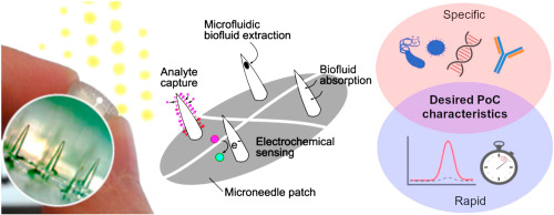
We have described the recent progress we have made in a novel drug delivery technology which, among other things, prolongs drug delivery beyond a month (read about it here , here and here). The research associate/assistant will be responsible for further development work to demonstrate a wide application of the drug delivery technology in various dosage forms, for the delivery of small molecules and macromolecular drugs (e.g. biologics).
This is a collaborative project between the School of Engineering and the School of Pharmacy at Newcastle University. Informal enquiries are welcome.