Ng KW, Archbold L, Lau WM. Building Otto: An open-source Franz diffusion cell autosampler for automating in vitro skin permeation studies. HardwareX. 2026;25:e00735. doi: 10.1016/j.ohx.2025.e00735
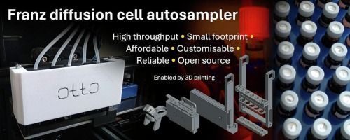
We have recently published a paper in HardwareX describing the design and construction of Otto, an open-source autosampler robot for Franz diffusion cell experiments. Otto is built using a desktop 3D printer as a gantry, a small number of custom 3D-printed parts, and commonly available laboratory consumables. It is designed to automate sampling and can collect up to 100 samples per run.
The system uses a Creality Ender 3 Pro 3D printer for motion control, with add-on components printed in-house on Prusa Research printers. The aim of the project was to develop a low-cost, accessible solution for automating repetitive sampling tasks in skin permeation studies, without reliance on proprietary hardware.
Otto has previously been validated. In a recent hydrogel study, Otto was used to collect every sample in a 72-hour skin permeation experiment, operating fully unattended throughout. The new HardwareX paper brings together the design rationale, build instructions and practical considerations needed for others to construct and use the system.
The paper, which is now available, provides a step-by-step guide to building Otto and is intended to support reproducibility and reuse by other laboratories. The 3D models and design files are openly available, and the models are also hosted on Printables.
Alongside the hardware, a companion application called OttoMate has been developed to generate the G-code used to control the system via a graphical user interface. The software is under active development and is available on GitHub.
A series of videos demonstrating different aspects of Otto’s operation, including assembly and sampling, is also available via a YouTube playlist.
This work was carried out by our team at Newcastle University, with contributions from Liam Archbold and Dr Wing Man Lau. Otto has been open-sourced in the hope that others will find it useful and adapt it for their own applications, and we are open to collaborations.
We would also like to acknowledge the EPSRC for funding this work.