Review: Silica nanoparticles in transmucosal drug delivery

Graphical abstract

Our latest review in drug delivery is about silica nanoparticles, published this week in the open access journal, Pharmaceutics.

This is a collaborative paper with Professor Vitaliy Khutoryanskiy (University of Reading, UK), Dr Twana M. Ways (University of Sulaimani, Iraq), and our own Dr Wing Man Lau (Newcastle University, UK). In the paper, we examine the applications of silica nanoparticles in transmucosal drug delivery. We discuss different types of silica nanoparticles and their methods of preparation, including surface functionalisation strategies to facilitate interactions with mucosal surfaces.

The paper is published under the very permissive Creative Commons Attribution Licence (CC BY 4.0), which allows you to freely share and adapt the work as long as the source is appropriately cited. Please cite this work as:

M. Ways TM, Ng KW, Lau WM, Khutoryanskiy VV (2020) Silica nanoparticles in transmucosal drug delivery. Pharmaceutics 12(8):751. doi: 10.3390/pharmaceutics12080751

Perspective: The diagnostic potential of microneedles in infectious diseases

The recent COVID-19 pandemic brought the healthcare systems in many countries to their knees. One of the key problems that became evident early on was the lack of diagnostic tools that were rapid and readily accessible to the public.

Diagnostics is what we do as a research team. Naturally, we contemplated what we could do in a situation like this. The result is a perspective article published in Precision Nanomedicine, the official journal of the European Foundation for Nanomedicine.

The conclusion? Microneedle systems excel in many ways as a diagnostic tool. Various microneedle platforms have demonstrated advantages of portability, self-administrability, affordability and scalability over other diagnostic platforms. The potential is huge but the technology is still in its infancy. We need more research to turn that potential into tangible clinical benefits, but we can’t do it alone. Cross-disciplinary collaboration and stakeholder support will be needed to drive this effort forward.

The article is published under the Creative Commons BY-NC-SA 4.0 licence, so please feel free to distribute widely, adapt and reuse for non-commercial purposes, and share any derived work, citing:

Dixon RV, Lau WM, Moghimi SM, Ng KW (2020) The diagnostic potential of microneedles in infectious diseases. Precision Nanomedicine 3(4):629–640 . https://doi.org/10.33218/001c.13658

Collaborative paper on 3D cell culture for evaluating biomarker-capturing microneedle devices

Update: Author manuscript available

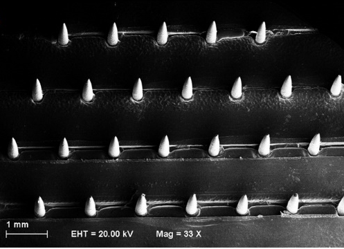
One of the challenges in developing microneedle devices to capture disease biomarkers from the skin is the lack of suitable skin specimens in which to test the devices. Donor skin specimens that carry the specific target diseases simply do not come by easily.

I first discussed this challenge with Eirini Velliou, Tao Chen and Guoping Lian in April 2016. We decided to tackle it by growing our own model of diseased skin in the lab. Experimental work started shortly after, and continued to develop following my relocation to Newcastle University in 2017. Earlier this week, we described the collaborative work in a joint publication in the journal Sensors and Actuators B: Chemical.

We took a grounds-up approach by growing melanoma skin cancer cells in a three-dimensional (3D) culture, supported structurally by a biocompatible polymer scaffold. This allowed us to simulate not only the biological microenvironment around the cells, but also the three-dimensional structure of the skin for microneedle insertion. Importantly, it was a simple and inexpensive, yet versatile, laboratory model to set up.

In pioneering the 3D cell culture model for evaluating microneedle devices against a skin cancer biomarker, we also demonstrated – for the first time – successful capture of S100B (a biomarker for melanoma skin cancer) in situ using our immunodiagnostic microneedle device.

Stella Totti, formerly a PhD student and now a postdoctoral researcher in Eirini’s research group, is the first author. I am especially pleased that hard work has paid off for Lorraine Dale, a former MSc student of mine, who contributed greatly to this work and is a co-author on this paper.

The paper is free to read until 28 July 2019 via this link: https://authors.elsevier.com/c/1ZBcy3IQMPEdJi

A microneedle array biosensor that can detect skin burns

SEM image of microneedle array biosensor

We have developed a microneedle array biosensor that can detect skin burns. To make the biosensor, we devised a nanocomposite material from carbon nanotubes and a biocompatible polymer, poly(lactic acid). We then shaped the microneedle array from the nanocomposite material. The microneedles that formed were about 870 μm long, 250 μm wide, and electroactive. This meant that the microneedle array could be inserted into  the skin to detect certain analytes by electrochemistry. We verified this by using the microneedle array to detect vitamin C in solution. Interestingly, when we tested the microneedle array biosensor on skin burns and normal (non-burnt) skin, the skin specimens showed different electrochemical responses. This gives us the technological basis for a minimally invasive biosensing approach to detecting skin burns.

For more information, please see:

Skaria E, et al. (2019) Poly(lactic) acid/carbon nanotube composite microneedle arrays for dermal biosensing. Analytical Chemistry (epub ahead of print). doi: 10.1021/acs.analchem.8b04980

Perspective: The future of skin biosensing and bioanalysis

I recently published a perspective article with Professor Syed Moein Moghimi in Precision Nanomedicine (the official journal of the European Foundation for Clinical Nanomedicine). In it, we offered our views and analysis on emerging techniques for sensing and analysing biomolecules in the skin, focusing on the capabilities and promises nanotechnology brings to the scene.

The article is open access.

In press: Nanoparticles designed to adhere to or penetrate mucosal tissue

Our paper on surface-functionalised nanoparticles and their ability to adhere to or penetrate mucous membranes has been accepted and published online. I congratulate my co-authors on this well-deserved outcome, and hereby present the paper to my readers:

MWays TM, Lau WM, Ng KW, Khutoryanskiy VV (2018) Synthesis of thiolated, PEGylated and POZylated silica nanoparticles and evaluation of their retention on rat intestinal mucosa in vitro. European Journal of Pharmaceutical Sciences 122:230-238.

The article is available for free from Elsevier until 29 August 2018. To access the free full text, please follow this link:

https://authors.elsevier.com/c/1XMgF3J1dIQyBp

In the paper, we demonstrated that nanoparticles whose surfaces were functionalised differently attained the ability to either adhere to or penetrate the mucus layer in the small intestines of the rat. We postulate that the same can be true in humans. This has important applications in oral drug delivery, where the nanoparticles can be tailored to target drug delivery to the gut differently, depending on the desired drug release profile.

The work was led by Professor Vitaliy Khutoryanskiy, of the University of Reading.

Hot off the press: high-throughput skin burn wound model for infection control

Graphical summary

A paper I co-authored with the team led by Dr Brian Jones (University of Bath) has just been published in the journal Frontiers in Cellular and Infection Microbiology. This is an open-access publication and you can grab a copy by following this link.

The paper describes a burn wound model that enables high-throughput evaluation of wound control measures. It is based on ex vivo (i.e. freshly excised) porcine skin that is both anatomically relevant and biologically responsive, and therefore superior to most in vitro models currently available.

I am glad to have contributed to this paper, and am pleased to introduce it to my readers.

Image credit

Editorial: Penetration enhancement of topical formulations

I have been working with the MDPI editorial office to guest edit the Penetration Enhancement of Topical Formulations special issue in the open-access journal, Pharmaceutics.

The collaboration has now concluded and I have written an editorial for that issue, which has now been published:

Ng, K.W. Penetration Enhancement of Topical Formulations. Pharmaceutics 2018, 10, 51

I thank the MDPI editorial office for the opportunity to be part of this effort. Special thanks go to MDPI editorial staffers, Felicia and Liane, who co-ordinated much of the work. It has been a most valuable experience for me, and I look forward to more future collaborations.