Climbing the Tree of Life

MartinIn this week’s blog, Professor Martin Embley reflects on the  the journey that led to him, his collaborators and his laboratory to fundamentally change our views on evolution and the Tree of Life.

The Early Years

My early career was a bit of a random walk while I tried to figure out what I really wanted to do. After my PhD in Newcastle on bacterial diseases of trout and salmon, I got a job teaching industrial microbiology at North East London Polytechnic in 1984. It was an odd but interesting place, a number of staff appeared to have strong religious beliefs of various sorts and wanted to talk about them, and one colleague thought he could change traffic lights from red to green so he never had to slow down. I was keen to keep doing some research and I was interested in evolution, but like a lot of newly independent researchers I struggled to get any funding. My big break came when I got a “cultural exchange” grant from Newham Council to go to Poland to learn some molecular biology and I met Professor Erko Stackebrandt who was passing through. Erko had worked with Carl Woese in pioneering the use of ribosomal RNA sequences to investigate evolutionary relationships among prokaryotes. I persuaded him to let me visit his lab in Germany to learn the new techniques and in 1991 these skills got me a job at the Natural History Museum in London.

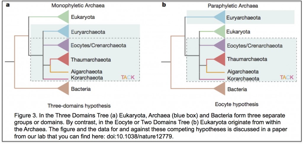
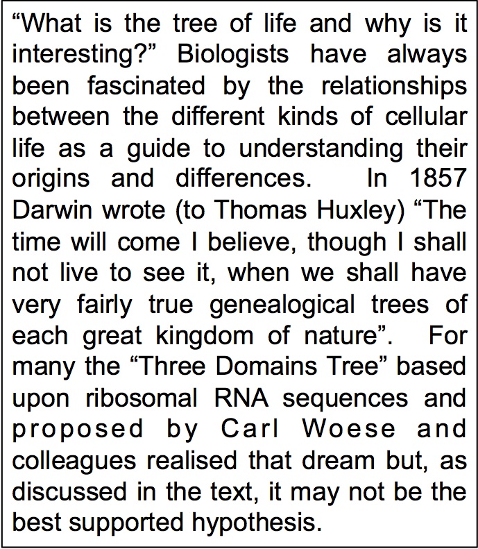
The Museum wanted to set up a lab using molecular sequences to investigate evolutionary relationships. The film Jurassic park was just about to appear and there was tremendous excitement about the potential of ancient DNA. The Museum gave me free rein regarding my own research as long as it had evolution at its core. So I decidedTree of life to work on the early evolution of eukaryotic cells. At the time two ideas were central to views of early eukaryotic evolution. One was that the “three domains tree of life” was an accurate description of the relationships between eukaryotes and prokaryotes (you can see the tree here). The other was that some eukaryotes, including obligate intracellular microsporidian pathogens, had never had mitochondria because they split from other eukaryotes before the mitochondrial endosymbiosis. I’ve been trying to test these two ideas for the past 25 years and while it’s often been difficult and frustrating, it has also been a lot of fun.
A Team Effort

Like most PI’s I’ve relied on attracting talented young scientists to do the work that we have published. Robert Hirt walked into my lab off the street and asked me if he could do a project involving eukaryotic evolution and ecology. He already had a first author paper in Cell and now he wanted to do something different. We didn’t do much ecologMitochondriay together but Robert and I co-supervised PhD student Bryony Williams who showed that microsporidians actually contained a tiny, hitherto overlooked mitochondrion, now often called a mitosome.
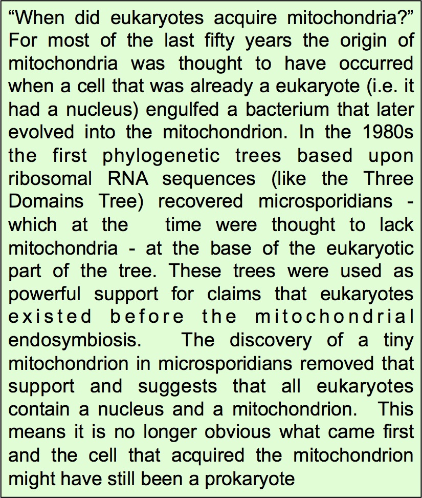
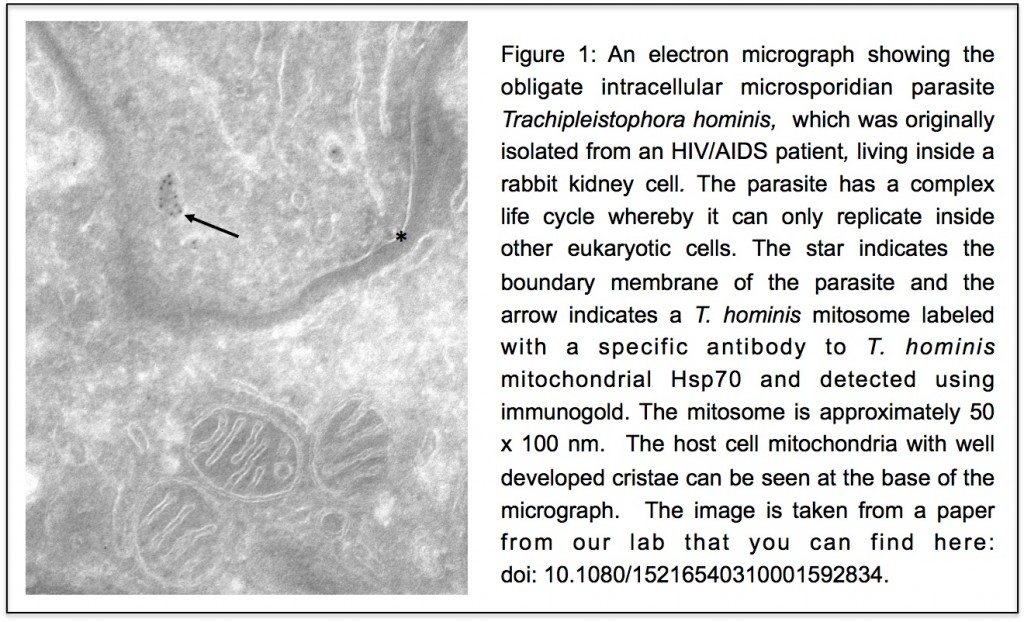
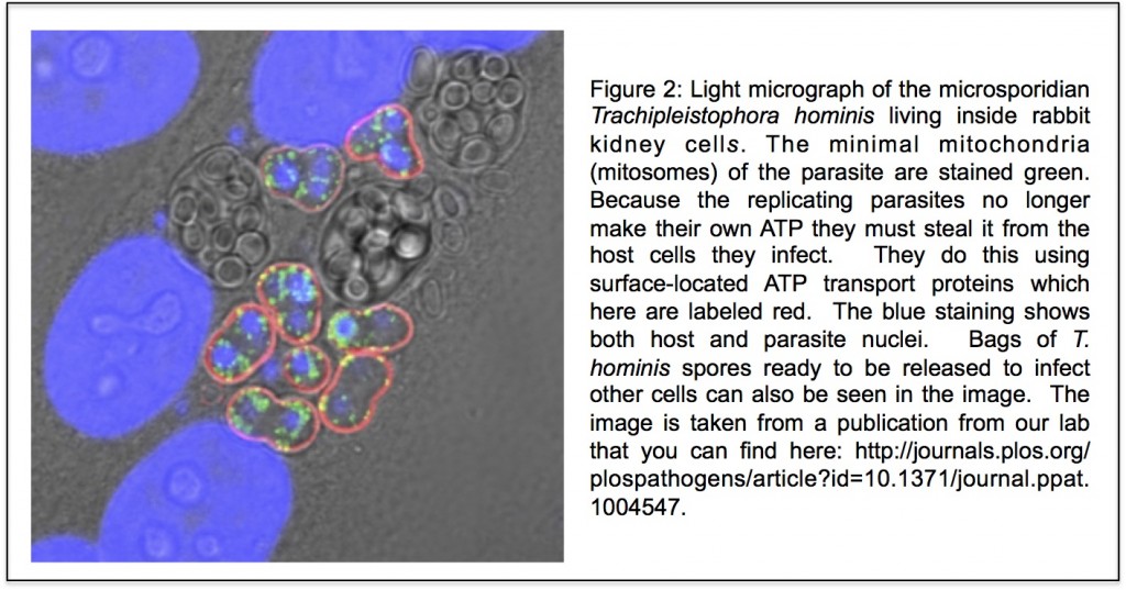
Unlike our own mitochondria, the microsporidian mitosome doesn’t make ATP, because it has lost all of the pathways used by classical mitochondria to make energy. Alina Goldberg in our lab – now at Newcastle – and Sabine Molik in the lab of Roland Lill in Germany spent Mitochondria 2the next seven years showing that the mitosome functions in the biosynthesis of essential cytosolic and nuclear Iron/Sulphur (Fe/S) proteins. The discovery of a tiny mitochondrion in microsporidia (Figures 1 and 2) was an important piece of evidence that led to current ideas that the mitochondrial endosymbiosis occurred at the origin of eukaryotes. Thus, it is now thought that all eukaryotes contain an organelle related to mitochondria, and its most conserved function is in Fe/S protein biogenesis, not ATP production.
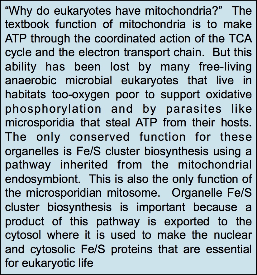
Figure 1Figure 2Competing Hypotheses

In the three domains tree of life, eukaryotes are a separate domain that is most closely related to the domain Archaea and the host for the mitochondrial endosymbiont is already a eukaryote. Although this hypothesis appears in most textbooks, there have actually been a number of alternative hypotheses published over the years (Figure 3 shows one of them), but these have largely been ignored. Cymon Cox spent three years analysing molecular sequence data to identify which of the competing published hypotheses was best supported and reached the surprising conclusion that it was not the three domains tree but an alternative hypothesis called the eocyte tree (you can read a discussion about the differences between the two trees including a picture of the eocyte tree here). In the eocyte tree, eukaryotes originate from within the Archaea, suggesting that eukaryotes are not a primary domain of life like Archaea and Bacteria, but are instead a product of genetic and cellular contributions from both prokaryote domains. Very excited by these results, we sent Cymon’s paper to Nature where it was reviewed and quickly rejected. We appealed, it was revised, it was reviewed again, and it was rejected again, all in all pretty dispiriting, but a common experience for most scientists. However, after hearing me talk about Cymon’s work at a meeting, we were invited to submit Cymon’s paper to PNAS where it was finally published in 2008.

Figure 3A Mixed Response

Although Cymon’s paper has been highly cited it is true to say that the initial response from the community was very mixed. We received emails suggesting that we had manipulated our results to get the answer we wanted and one of the reviewers told us that it was impossible to infer such ancient events using molecular sequences. In responding we agreed that it was difficult to be confident about anything that happened billions of years ago based upon small amounts of data and even the best methods of analysis, but that people in the field seemed happy to use the same data and worse methods to support the three domains tree. Cymon eventually moved on, scarred but not defeated, and Tom Williams took over the project when he came to Newcastle on a Marie Curie Fellowship in late 2010. Over the next few years, more data was published as new Archaea were discovered and new methods of analysis were developed, and every analysis that Tom, or others, did on these data produced a version of the eocyte tree, so that it is now the best supported hypothesis – at least in our opinion.

More evidence emerges

Hypotheses are only useful when they make predictions that can be tested by further research, and evolutionary hypotheses are no different. The eocyte tree predicts that new species that share more features in common with eukaryotes will be discovered among the Archaea, and this prediction now appears to have been spectacularly fulfilled by recent discoveries from Thijs Ettema’s lab in Sweden. The new paper describes the discovery, so far only from metagenome data, of an archaeal lineage called Lokiarchaeota that contains many genes for proteins that were previously thought to be eukaryotic specific, including homologues of proteins used in the eukaryotic cytoskeleton, in membrane remodeling and in phagocytosis.  This is incredibly exciting and the challenge is now to isolate Lokiarchaeota and other new lineages into culture so that their biology and physiology can be studied in the laboratory.

An Interesting Journey

Scientific work is often written up as if it were a linear progression towards improved understanding, a type of “Whig history” which does not accurately reflect how science is really done. In reality, science is a collaborative endeavour with lots of dead ends, confusion and false trails, and we could easily be walking down some of those still. Nevertheless, the currently prevailing paradigm for eukaryotic evolution is now very different to the popular views held in the 1990s when I started my research career. All eukaryotes are now thought to contain a mitochondrial homologue that generally functions in Fe/S protein biogenesis, and the host for the mitochondrial endosymbiont is thought to have originated from within the Archaea. Eukaryotes are thus viewed as the product of an interaction between (at least) those two prokaryotic partners and are not a primary domain of life but one derived from prokaryotic antecedents. The complex features that we take to define eukaryotic cells including our own, such as the nucleus, large genomes and diversity of RNAs, are thus secondary features that have evolved since those primordial interactions. I’m not sure what my religious former colleagues would have made of the work I’ve done since leaving NELP, but it’s been an enjoyable and interesting journey for me.

Links

The Tree of Life: http://www.pnas.org/content/87/12/4576.full.pdf

Bryony Williams’ paper: http://www.nature.com/nature/journal/v418/n6900/full/nature00949.html

Alina Goldberg and Saline Molik paper: http://www.nature.com/nature/journal/v452/n7187/full/nature06606.html

Mitochondria and Fe/S proteins: http://www.nature.com/nature/journal/v440/n7084/abs/nature04546.html

The eocyte tree: http://phenomena.nationalgeographic.com/2012/12/20/redrawing-the-tree-of-life/

Cymon Cox paper: http://www.pnas.org/content/105/51/20356.full

Tom WIlliams paper: http://www.nature.com/nature/journal/v504/n7479/full/nature12779.html

Thijs Ettema lab paper: http://www.nature.com/nature/journal/v521/n7551/abs/nature14447.html

Donating hope – a success for parents in danger of transmitting mitochondrial disease

 by Prof Bob Lightowlers

It was 2pm on a Tuesday afternoon and there I was watching television in my office. An unusual experience (honestly) but it was to be a truly momentous occasion. For the next 90 minutes, members of parliament would be debating whether to sanction the procedure of mitochondrial donation. What on earth does that mean ? Well, mitochondria are essential compartments (or organelles) that provide numerous key functions for the cell. They also have their own genome, called mitochondrial DNA – mtDNA – that contains the information to make up just 13 proteins, all of which are important in their function of producing main energy source of the cell, ATP. This is why mitochondria are often referred to as the cell’s batteries or the powerhouse of the cell. It is important to note that mtDNA is very small when compared to the nuclear DNA component: 16 thousand mitochondrial nucleotides (that is, the “letters” in the genetic code) vs more than 3 billion nucleotides in the nuclear DNA.

OK, but why would you want to donate mitochondria?

Mitochondrial DNA is strictly passed down via our mothers, unlike nuclear DNA which is comes from our father and mother. Almost 2,500 women in the UK have mutations in some or all of their mtDNA that can cause disease. Defects of this mitochondrial genome, can be responsible for a wide spectrum of mainly muscle and neurodegenerative disorders for which there is no treatment. By identifying those women with defective mtDNA, we could potentially prevent transmission of their unhealthy DNA by substituting their mitochondria for organelles from a donor. Simple on paper!

What’s the problem?

There are two major barriers. First, how safe would any technique be for mitochondrial donation ?

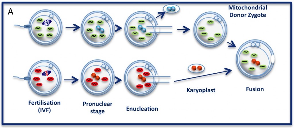
The pronuclear transfer method: Embryos are shown with mitochondria carrying normal (green) or mutant (red) mtDNA. As the embryos begin to develop, pronuclei become visible. Pronuclei from the normal donor embryo are removed (blue, top panel ‘enucleation’) and are replaced with the nuclear  DNA from the patients (red, karyoplast). The resultant embryo carries nuclear DNA from the patients and mtDNA from the donor (mitochondrial donor zygote).

The pronuclear transfer method: Embryos are shown with mitochondria carrying normal (green) or mutant (red) mtDNA.
As the embryos begin to develop, pronuclei become visible. Pronuclei from the normal donor embryo are removed (blue, top panel ‘enucleation’) and are replaced with the nuclear DNA from the patients (red, karyoplast). The resultant embryo carries nuclear DNA from the patients and mtDNA from the donor (mitochondrial donor zygote).

The technique being championed in Newcastle is that of pronuclear transfer. The idea is to take the nuclear DNA from a fertilised embryo and transfer the DNA to a donor with no nuclear DNA, ie, where the nucleus was removed. The newly made embryo then has nuclear DNA from the mother and father but has only mtDNA from the donor.
As you can tell, there is quite a lot of tricky manipulation here. Further, although the mtDNA that has been replaced carries only a very small number of genes, could these somehow be incompatible with the nuclear DNA?
After many years of very careful analysis, there is no evidence to suggest that this procedure is unsafe. The question of incompatibility would also seem to be highly unlikely. After all, nature has been performing the experiment of mixing and matching nuclear and mtDNA since the evolution of Homo sapiens. The idea that, for example, there would be something wrong with the child of an aboriginal woman and a inuit man due to the substantial differences in their mitochondrial DNA would seem laughable.

In addition, does the replacement of DNA, albeit the complete mitochondrial genome, constitute genetic manipulation? This is a contentious issue and a strict definition of what constitutes genetic manipulation, particularly when concerning mtDNA, is difficult to agree on. It must be remembered that mtDNA is completely separate from nuclear DNA and needs to be considered as such.

A separate issue is that many people are ethically uncomfortable with this process. Can embryo manipulation ever be acceptable? Certainly, many religious people have a deeply felt objection to this.
Even when we accept that these methods could offer such an immeasurable benefit for many couples, it is clear that there were many questions to be tackled before we could consider the prospect of mitochondrial donation. For this reason, experimentation and public consultations had to be initiated.

The Parliamentary Under-Secretary of State for Health, Jane Ellison, when presenting the vote on Tuesday highlighted the measures taken to date to assess the safety and ethical concerns surrounding mitochondrial donation.

The Parliamentary Under-Secretary of State for Health, Jane Ellison, when presenting the vote on Tuesday highlighted the measures taken to date to assess the safety and ethical concerns surrounding mitochondrial donation.

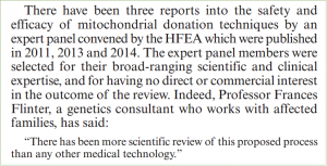
To cut a very long story short, both have been carried out for many years, including supportive public consultations and independent review by the Human Fertilisation and Embryology Authority (HFEA) reporting that the procedure was not dangerous, as we’ve covered before (here and here). Following these procedures, Professors Turnbull, Herbert and numerous members of the Wellcome Trust Centre for Mitochondrial Research were instrumental in persuading the government to finally hold a debate in the House of Commons.

The vote scheduled for Tuesday afternoon would decide whether the HFEA would have the right to offer a licence to perform mitochondrial donation, a first for the UK and the world. The week leading up to the debate was a white-knuckle ride. Letters of support were published in leading newspapers from Nobel Laureates and other eminent scientists. Just when it appeared that the tide was turning, the Church of England announced it could not support mitochondrial donation. This was a great disappointment and rather a shock, as they had been involved throughout the lengthy consultation processes and had not indicated their level of concern.

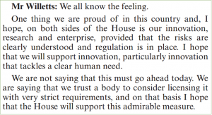
Now a back-benh MP, former Minister for Science, David Willets, made a clear case in support of mitochondrial donation.

Now a back-bench MP, former Minister for Science, David Willetts, made a clear case in support of mitochondrial donation. You can read the whole debate here.

Back to me watching the television. At 3:45pm, the members had cast their votes and the count was in – 382 for the motion, 128 against! This is a fantastic result. It gives couples who may be at risk of having a baby with mitochondrial disease the chance to choose whether they want to try mitochondrial donation, just like couples have been able to choose in vitro fertilisation since the 1970’s.

Obviously, this result has gathered lots of media interest and even I was rolled out to perform a couple of interviews.

There is still more to be done, however. Further important research to support the safety of the procedure is currently in review but it must be recognised that every clinical procedure carries a risk. What this vote does is to empower the HFEA to licence this procedure in the UK, but this is still an important barrier and many issues are still to be addressed. And it also needs to be discussed by the House of Lords, of course.

Regardless, today’s vote was a wonderful day for anyone who has been touched by mitochondrial disease in any form. Twenty years ago, Doug Turnbull and I used to discuss this idea. He and his colleagues have done a remarkable job to make this pipedream a reality.

Breaking news: Mitochondria replacement therapy given green light

Breaking news by Prof Bob Lightowlers

Professor Dame Sally Davies, the Governments Chief Medical Officer, has today given support to a change in the law that will allow the form of mitochondrial replacement therapy that is being pioneered here in Newcastle, has we mentioned before.

Its great news for any prospective mother with mitochondrial DNA disease who is concerned about transmitting the disease to their children.

Most media is covering this – BBC, Guardian, Telegraph – but if you are still confused, we thought it would be good to recap the essential points:

Electron micrograph of a cell (coloured blue) revealing part of the mitochondrial structure (orange) within. The entire length of the mitochondrion is about 5 micrometres.

– Mitochondria are crucial structures found in all cells in our body and they take our common foodstuffs such as fats and sugars and turn them into energy. They have their own genetic element, mitochondrial (mt) DNA. Much smaller than our chromosomes, mtDNA is essential for energy production.

– In 1988, scientists in the UK and US recognised that certain diseases were caused by mutations in mtDNA with the main disorders related to your muscle tissue and the brain.

– It is estimated that at least 1:10,000 people suffer from disorders associated with defects in mtDNA – that’s more than 6,000 people in the UK.

– Mitochondrial DNA is only transmitted to babies by their mothers. Unfortunately, as you inherit your mothers mitochondria, diseases caused by mtDNA mutations are inadvertently transmitted from the mother.

– Doug Turnbull, Director of the Wellcome Trust Centre for Mitochondrial Research (WTCMR) and I have wondered since the 1990s whether it would be possible to prevent the transmission of the faulty mtDNA from the mothers to their children by transferring the nucleous from an egg of the mother carrying these mtDNA mutations into a healthy egg whose nucleous had been removed, and then fertilise it

– Professor Mary Herbert, Doug and a group of us from the Mitochondrial Research Group were able to show that such a swap could be performed without any or very low levels of the defective mtDNA being transferred. Importantly, there was also no defect detectable in the reconstituted cells.

In August 2012, the government asked the Human Fertility and Embryological Authority (HFEA) to find out what the general public thought of the procedure.  The results were collated  and the Human Fertility and Embryological Authority made a recommendation to Government. There was an overall support for the new technology with only 10% being fairly or strongly against the concept of mitochondrial gene replacement

To understand all the details and find out the whole story, see my post a while ago here.

The Wellcome Trust also has a very nice video explaining the science behind the news story:


Links

Wellcome Trust Centre for Mitochondrial Research http://www.newcastle-mitochondria.com/
Human Fertility and Embryological Authority (HFEA) http://www.hfea.gov.uk/index.html
HFEA mitochondria puclib consultation 2012 http://www.hfea.gov.uk/6896.html


You don’t always want what your mother gives you! – can we prevent mitochondrial disease?


By Professor Robert Lightowlers

In 1988, scientists in the UK and US recognised that certain diseases were caused by mutations in mtDNA . Over the following 20 years, mtDNA defects have been shown to cause a range of debilitating diseases many affecting different parts of the body. However, the main disorders relate to your muscle tissue and the brain.

Human muscle fibres stained for mitochondrial function. As can be seen in B, some of the fibres show no activity. This is because these fibres have high levels of mutated mitochondrial DNA.

It is estimated that at least 1:10,000 people suffer from disorders associated with defects in Mitochondrial DNA (mtDNA) – that’s more than 6,000 people in the UK. Even so, it is only recently that the importance of mitochondrial diseases have hit the general media.

Many of you will have seen the debate on correcting mitochondrial diseases in the newspapers (for example, see the Guardian, Telegraph) and on television recently, but not be aware of the central role that Newcastle researchers have played in making this exciting, or to some, controversial, new therapy closer to becoming a reality.  Here, Bob Lightowlers ICAMB Director and senior member of the Wellcome Trust Centre for Mitochondrial Research (WTCMR) reflects on the role mitochondrial research in Newcastle has played in this process over the last 20 years and tells us some of the story behind the headlines.

What are Mitochondria?

Electron micrograph of a cell (coloured blue) revealing part of the mitochondrial structure (orange) within. The entire length of the mitochondrion is about 5 micrometres.


These crucial structures found in all the trillions of cells in our body have many essential functions. One very important role they play is to take our common foodstuffs such as fats and sugars and turn them into energy for our body’s to function.

A single human cell showing the nucleus (green), the mitochondrial network (red) and the mitochondrial DNA within the network (yellow)


One surprising element of these structures is that they contain their own genetic element, mitochondrial (mt) DNA. Much smaller than our chromosomes, mtDNA is essential for energy production.


OK, so this is important, but why have mitochondria and mtDNA begun to work their way into the common conversation of the nation?

Answer: Our mothers!

What has this got to do with our mothers ? Mitochondrial DNA is only transmitted to babies by their mothers. This is different to all our other DNA where copies are made and transmitted from both parents. Unfortunately, as you inherit your mothers mitochondria, diseases caused by mtDNA mutations are inadvertently transmitted from the mother.

How does this relate to Newcastle based Mitochondrial Research?

My colleague Doug Turnbull, a neurologist here in Newcastle (and Director of the WTCMR) and I have been intrigued by these mtDNA mutations since it first became clear that they could cause disease. Back in the early ‘90’s, we discussed whether some day it would be possible to try and prevent the transmission of the faulty mtDNA from the mothers to their children. Of course, at that stage, it was just wishful thinking. As the Mitochondrial Research Group (MRG) began to grow and mature in Newcastle, we often returned to one question:

What if the nucleus from the diseased egg could be transferred to a healthy egg whose nucleus had been removed, in essence leaving all the affected mtDNA behind ?

If it was indeed possible, this reconstituted egg could be fertilised and implanted back into the mother by standard techniques used routinely in fertility clinics throughout the world. We also would consider when would such a technique be most efficient: before or after fertilisation of the egg? On paper both options looked possible, but there are many complications.

Technical Concept: achieving the switch of nuclei without some of the faulty mtDNA being inadvertently taken along for the ride.

Towards the end of the 90’s, scientists working in Canada were able to show that the level of mtDNA inadvertently transferred when the nucleus was switched into a recipient cell lacking a nucleus, was low. This was a promising result, but it led to two central questions:

•   Could this be repeated with human cells?

•   Was this technique morally and ethically acceptable to everyone?

The ethical debate: Debate raged as to whether this technique would constitute genetic manipulation of humans, which of course would be illegal. Further, it was not possible to perform these types of reconstruction experiments in man, as using viable human fertilised cells for research was also, understandably, illegal.

Professor Mary Herbert working at the nearby Human Fertility Centre came up with an intriguing proposal. She explained that unfortunately, during the standard process of in vitro fertilisation, many eggs became incorrectly fertilised. These eggs are unable to grow correctly and have to be discarded. One way of determining whether it would be possible to swap mtDNA in humans, she suggested, was to use these incorrectly fertilised eggs. As this procedure would still require the manipulation of fertilised human eggs, a licence would need to be applied for from the Human Fertilisation and Embryological Authority (HFEA). Following lengthy and extensive debate, including members of the research team being called to the House of Commons, a licence was eventually awarded in 2005.  Five years later, with the essential help of colleagues in the Fertility Centre, Mary, Doug and a group of us from the MRG were able to show that such a swap could be performed without any or very low levels of the defective mtDNA being transferred. Importantly, there was also no defect detectable in the reconstituted cells . In 2011, this very promising result, along with many other important contributions made by the Newcastle MRG to understanding mitochondrial biology in health and disease was recognised by the Wellcome Trust who funded the establishment of a new Research Centre in Newcastle, the Wellcome Trust Centre for Mitochondrial Research.

Getting acceptance of the technique

It was important to know whether the people of the UK agreed that such reconstitution technology was ethically acceptable. In August 2012, the government asked the Human Fertility and Embryological Authority (HFEA) to find out what the general public thought of the procedure .  The results were collated last month and the Human Fertility and Embryological Authority made a recommendation to Government. There was an overall support for the new technology with only 10% being fairly or strongly against the concept of mitochondrial gene replacement ) This is an endorsement of the method but there is still a long way to go before the technique can be performed in the clinic.

Its amazing to think how far this concept has come in 20 years. Perhaps in another 20 years we may be able to look back and celebrate how this dream has helped to provide a realistic method to help prevent the transmission of a debilitating disease for many couples.


Wellcome Trust Centre for Mitochondrial Research http://www.newcastle-mitochondria.com/
Human Fertility and Embryological Authority (HFEA) http://www.hfea.gov.uk/index.html
HFEA mitochondria puclib consultation 2012 http://www.hfea.gov.uk/6896.html