Determination is key – Prof Ramakrishnan’s Baddiley Lecture

By Kevin Waldron.

Last week saw ICaMB host the latest in our series of Baddiley lectures, which commemorates Professor Sir James Baddiley (1918-2008). Baddiley was a distinguished Professor at Newcastle University (1954-81) and a Fellow of the Royal Society (elected 1961) who made numerous important fundamental discoveries in microbiology, not least the discovery of teichoic acids, cell wall components in Gram positive bacteria.

Jeff Errington introduces Venki Ramakrishnan to the ICaMB audience

Jeff Errington introduces Venki Ramakrishnan to the ICaMB audience

Baddiley’s work on the fundamental processes of bacteria, including the structure and function of components of the bacterial cell wall, is continued to this day in Newcastle through the work of members of ICaMB’s Centre for Bacterial Cell Biology (CBCB). Although James Baddiley died shortly after the first in ICaMB’s series of Baddiley lectures, we were delighted that the Baddiley family was again represented at this year’s lecture by James’s son, Christopher Baddiley.

This year’s guest speaker was Professor Sir Venki Ramakrishnan, distinguished research leader and Deputy Director of the Laboratory of Molecular Biology in Cambridge, Nobel laureate and newly-elected President of the Royal Society. With all of the demands on his time that come with this new role as President, the large audience that gathered on Friday afternoon were grateful that Venki was able to find time to visit Newcastle to deliver his lecture. As ever, both the lecture and the surrounding celebration was expertly organised and introduced by CBCB Director, Professor Jeff Errington.

Venki illustrates the structure of the yeast mitochondrial ribosome

Venki illustrates the structure of the yeast mitochondrial ribosome

Venki’s lecture gave a brief history of his atomic-resolution structural studies of the ribosome, the macromolecular nucleoprotein complex that converts the four-letter genetic code in nucleic acid into the twenty-letter amino acid code in proteins. He presented detailed structural models of eukaryotic ribosomes, derived from X-ray crystallography and cryo-electron microscopy data accumulated over 30 years of detailed study in his laboratory.

We asked some of ICaMB’s early career researchers to describe their impression of Venki’s lecture:

“Professor Venki Ramakrishnan was kind enough to deliver this year’s Baddiley lecture. It was an honour to meet Venki, who somehow managed to fit us in between Royal Society committee meetings and a chat with the Science minister! He impressed us all with a phenomenal talk discussing how he solved the structure of the mitochondrial ribosome using cryo electron microscopy. Wow – cryo EM has truly moved beyond blob-ology! One thing that really struck me about Venki is how humble he is; despite being so incredibly successful and lauded, there’s not a trace of ego on the guy; something for us all to aspire to.”

Venki illustrates how the ribosome works

Venki illustrates how the ribosome works

Seamus Holden, University Research Fellow

“It was incredibly cool to hear about Venki’s work first hand. The enormity of his achievement became clear when he showed a single slide with the dozens of conformations of the ribosome’s catalytic cycle and indicated that there were structures available for the majority of them! And what was humbling was that Venki did not seem at all interested in dwelling upon his past successes. Rather, he briskly moved past this slide onto his current work regarding mitochondrial ribosomes which was both cutting-edge but also somewhat raw because of its novelty. I found it inspiring to see a scientist of his stature still so driven to continue discovering and learning.”

Post lecture, Venki holds the gift he received from his hosts at ICaMB

Post lecture, Venki holds the gift he received from his hosts at ICaMB

Heath Murray, Royal Society URF

“For me Venki’s journey was an excellent advert for never giving up. Often as researchers (particularly at the start of our careers) we are encouraged to know when to call time on a set of experiments that are bogged down and not yielding answers. Pursuit of the next grant and the speed of some of our competitors unfortunately make it risky to continue to spend years and years believing in the same project that fails to show progress relatively quickly. Yet what an example Venki is, many years and many postdocs focusing on the same problem, persistence and belief in himself and his team has more than paid off. Inspirational.”

Suzanne Madgwick, Wellcome Trust Career Re-entry Fellow

Congratulations, Prof Gilbert, FRS!

by Prof. Harry Gilbert, FRS, FMedSci

I was asked to write a blog about my election as a Fellow of the Royal Society. I start by apologising for my ineptitude compared to the “professional” social media people in the Institute. So, what to say? Well, maybe the process might be of interest.

How do you get elected as an FRS?

ICaMB's FRS: Prof Jeff Errington and Prof Harry Gilbert

ICaMB’s FRS: Prof Jeff Errington and Prof Harry Gilbert

You need to be proposed and seconded by current FRS’s. Jeff tried twice to propose me before I went to the USA and a third time (September 2012) upon my return, at which point I said OK. I had to generate a full CV, list 20 papers and include PDF versions of these articles, and the proposers were required to write a three page narrative on my research.

 
Every year material is updated. The first year I wondered what feedback I might get but I soon realized that no one says anything to you. I rapidly put the issue to one side and did not give it any thought.

miebio2014570

‘Wrong Direction’ – Prof Gilbert forms a karaoke boyband with other eminent scientists at a conference in Japan


Looking back on my research it’s evident that most of my science was done with collaborators who are much smarter than me. So, I consider myself to be extremely fortunate to have worked with these people.


The talented people currently working in the Gilbert/Bolam labs

The talented people currently working in the Gilbert/Bolam labs

Bernard_quote

Finally, the election

In late March of this year, I received a letter from the Royal Society marked “In strictest confidence“. I assumed it was yet another reference for someone applying for a grant to the Royal Society and I thought “another job to do”. I read the letter several times and the words “you are on the list of candidates submitted to become an FRS” was hard to digest. When it said you needed 2/3 of the votes to be elected, I became dubious about my chances. On speaking to Jeff, he explained that only one person in the last 150 years has failed to become an FRS at this stage. I was thus confident that not even I could screw this up.

Fun celebrations!

Fun celebrations!

However, keeping it secret for a month before the election took place was extremely hard as I don’t do secrets. I did, however, tell Rosie, my wife, who initially shared my excitement of the news. Her enthusiasm waned somewhat as I kept on about it at home for a day or two. At this point, Rosie said “I hope you don’t become too grand” which ceased any talk about the FRS. I am pleased to say that Rosie’s excitement was rekindled when she realised that she will likely get the opportunity to meet Brian Cox (who also became an FRS this year) who she rates as “very dishy, particularly for a scientist”.


How does it feel to be an FRS?

What are my thoughts about being an FRS? Well, shocked but also excited, although I feel a complete fraud.

Prof Gilbert's speech to his colleagues at ICaMB

Prof Gilbert’s speech to his colleagues at ICaMB

I was very pleased that so many people were able to come and celebrate with me on Friday, I would like to thank you all for coming. I also hope that people will think that “if Harry can become an FRS then the bar is not so high”, and that this will result in other people becoming Fellows in the next few years. In 30 years at Newcastle, I have never worked in a place with so many talented people, and it is clear that ICaMB merits more than two Fellows of the Royal Society.

bobquote2

ECRs at ICaMB: RNA Quality Control


Claudia SchneiderIn the latest of our series focussing on the ECRs in ICaMB, we feature Dr Claudia Schneider. Claudia obtained her PhD from the Philipps-University in Marburg, Germany. She then moved to the UK to work with Prof David Tollervey at the Wellcome Trust Centre for Cell Biology in Edinburgh. In 2011, she was awarded a Royal Society University Research Fellowship and started her own lab at ICaMB. Here, Claudia describes her research, and how being alarmed during her postgraduate studies triggered her long-term research interest.

By Dr Claudia Schneider

Hi, my name is Claudia Schneider, and my Royal Society University Research Fellowship has allowed me to set up my own group here at ICaMB to study enzymes involved in RNA processing and quality control.

I am originally from Germany, where I did my undergraduate studies and my PhD. Many people might think that Germany is the land of cars or lederhosen – but in truth it is really the land of bread (and beer!). It might therefore not come as a surprise that baker’s yeast has become my favourite model organism.

Click on the image to find out how to make these budding buns!

Click on the image to find out how to make these budding buns!

During my undergraduate studies I was first introduced to RNA and I was (and am still) amazed by its many known and still emerging functions in the cell. We now know that almost the entire eukaryotic genome is transcribed, but only a small fraction of the transcripts are protein-coding messenger RNAs (mRNAs). The others are stable and unstable non-coding RNAs (ncRNAs), which are involved in all aspects of gene expression. RNA molecules are often extensively processed before they’re functional, and each processing step is subject to quality control mechanisms. If you want to know more about the life and death of non-coding RNAs, have a look at this recent review.

Yeast has not always been my first choice to study RNA metabolism, since the object of my PhD project in Prof Reinhard Lührmann’s lab turned out to be completely missing in baker’s yeast. Back then I worked on nuclear pre-mRNA splicing, the removal of non-coding introns from precursor mRNAs catalysed by the spliceosome. It was an exciting time in the splicing field: A second low abundance “minor” spliceosome had just been discovered in most multicellular eukaryotes (with the strange and still not readily explainable exception of C. elegans), and this complex is not present in yeast. The minor spliceosome recognises a rare class of introns (<0.5%) with different consensus sequences at the splice sites and has since been linked to a number of human diseases. During my PhD, I purified and biochemically characterised the snRNP components of this unusual pre-mRNA splicing machinery in human and Drosophila cells.

Since then, I have been fascinated by biochemical and enzymatic assays involving RNA such as in vitro splicing assays, where in vitro transcribed pre-mRNAs are mixed with purified spliceosomes. The goal of such an experiment is to observe precise and (hopefully) pretty intron removal in the test tube – but, to my great annoyance, success was every so often hampered by a powerful ribonuclease (RNase) contamination in the assay that completely trashed the precious RNA substrates. Generic and aggressive RNases like RNase A are found on our skin, are incredibly stable and can even survive boiling.

Common decoration on lab surfaces during my PhD

Common decoration on lab surfaces during my PhD

It is therefore fair to say that my scientific career was majorly influenced by constant warnings by my mentor, who told me that all RNases are evil and must be destroyed. However, for my postdoc, I decided to face my fears and look these evil RNases in the eye, in the humble model system yeast. During my time with Prof David Tollervey at the University of Edinburgh, I learned that there are many different types of RNases, and only very few of them are promiscuous and chop RNA to bits.

The majority of RNases are very sophisticated and versatile enzymes. Several RNases are capable of degrading only specific RNA molecules, or only function under certain circumstances, and protein co-factors often assist in substrate recognition. RNases are crucial elements in RNA quality control or surveillance systems, which distinguish aberrant from “normal” RNA molecules. One clinically important RNA surveillance pathway is called “nonsense-mediated decay” or NMD, and this system recognises and degrades a specific class of defective mRNAs to limit the synthesis of truncated and potentially toxic proteins. NMD defects are linked to ~30% of all inherited human diseases (e.g. Duchenne muscular dystrophy and forms of b-thalassemia) as well as certain types of cancer. In addition to quality control/surveillance, where RNAs are mostly completely degraded, a growing number of RNases have been shown to be responsible for precise processing or “trimming” of precursor RNA molecules to produce their functional forms.

Exonucleases were long believed to be the main players involved in RNA recognition and processing/turnover. However, this model was recently challenged by the identification of endonucleases containing PIN (PilT N-terminus) domains, which appear to play key roles in RNA metabolism. Eight PIN domain proteins and therefore putative endonucleases are encoded in the genome of budding yeast, and this includes three largely uncharacterised “orphan” nucleases.

Overall it is still puzzling to me how individual RNases “make the decision” to either completely degrade or carefully process a specific RNA. Given the ever-growing number of non-coding transcripts in the cell, I am also keen to know which RNases are responsible for which substrates and what the so-far uncharacterised putative PIN domain endonucleases in yeast are doing!

To this end, our lab is using an RNA-protein cross-linking method called “CRAC” (UV cross-linking and analysis of cDNA) to identify the targets of PIN domain endonucleases on a transcriptome-wide scale. The CRAC method and the machinery to cross-link yeast cultures were developed by Sander Granneman at the University of Edinburgh, when we were both PostDocs in Prof David Tollervey’s lab. Sander now has his own lab too, and he runs a CRAC-blog. Interestingly, the cross-linking device we are using was originally designed to sterilise sewage water, but it is now also commercially available for research. With this setup, yeast cells are cross-linked while they are growing in culture, which is crucial to identify the often very transient interactions between nucleases and their target RNAs. This system provides a huge advantage over more traditional cross-linkers like the “Stratalinker”, which requires pelleting and cooling the cells on ice before cross-linking. It is, however, also much bigger and takes up a whole bench in the lab – but I guess there is a drawback to everything! In any case: if you want to find RNA targets for your favourite yeast protein – get in touch!!

Cultures of Saccharomyces cerevisiae can be “zapped”, while they are growing: It only takes 100 seconds!

Cultures of Saccharomyces cerevisiae can be “zapped”, while they are growing: It only takes 100 seconds!

Transcriptome-wide RNA-protein interaction analyses generate huge datasets and we use RNA binding and nuclease assays, as well as co-precipitation studies, to validate the in vivo cross-linking results for individual PIN domain endonucleases.

With the help of an ERASMUS exchange student, Franziska Weichmann, who spent 6 months in my lab last year, we have made good progress with two putative PIN domain endonucleases that are linked to ribosome biogenesis. We were able to identify their binding sites on the pre-ribosomal RNAs, as well as co-factors that are important to recruit them into the pre-ribosome. We have also set up an in vitro system with recombinant proteins, and we are currently trying to convince one of them to specifically cleave its proposed rRNA substrate in the test tube – and we are slowly getting there…..

Like the other ICaMB ECRs, who posted on this Blog before, I would like to finish by saying that having my own lab has been an exciting and (on most days) enjoyable adventure so far – and I am looking forward to the next set of challenges…

ECRs at ICaMB: Copying the blueprint of life – Understanding DNA replication


Heath MurrayIn the latest of our series focussing on the Early Career Researchers (ECRs) in ICaMB, we feature Dr Heath Murray.  After completing undergraduate studies at the University of California, Los Angeles and then obtaining his Ph.D. from the University of Wisconsin-Madison, Heath came to the UK to join the lab of Prof Jeff Errington in Oxford. From there he re-located to ICaMB, and in 2009 was awarded a Royal Society University Research Fellowship. Here, Heath describes his research into the mechanisms of DNA replication, and explains why he became interested in this field.

By Dr Heath Murray

Hello, my name is Heath Murray and I’m a Royal Society University Research Fellow in ICaMB’s Centre for Bacterial Cell Biology (CBCB) studying DNA replication. DNA is one of the most important molecules required for life because it encodes the information, or the blueprint, used to build a cell (i.e. the most basic unit of an organism). In order for a cell to create new cells it must synthesize an exact copy of its DNA, an extraordinary process when you consider that the genomes of most cells contain millions of individual DNA subunits!

Bacillus subtilis is a useful model system as it proliferates rapidly and is amenable to genetic, cell biological, biochemical, and structural analyses

Bacillus subtilis is a useful model system as it proliferates rapidly and is amenable to genetic, cell biological, biochemical, and structural analyses

Bacteria are ideal model systems to study this fundamental process because they are much less complex than human cells (e.g. all of their DNA is encoded by a single chromosome, whereas humans have 23), and this allows us to understand how they work at the greatest possible level of detail.

I was introduced to bacteria when I was an undergraduate student and the effect was transformative. My mentors taught me how to add a specific gene (a DNA sequence) to a bacterial cell, and if it worked properly then the bacteria would turn blue!

Bacterial colonies turn blue if they contain a gene that degrades specific sugars.

Bacterial colonies turn blue if they contain a gene that degrades specific sugars.

That basic genetic experiment was one of the coolest things I had ever done, and from that point on I worked hard to learn the trade of “bacterial genetics”.

Today my research group focuses on understanding how DNA replication is controlled so that each new cell will end up containing an exact copy of the genetic material from its predecessor. We employ a wide range of complementary experimental techniques: genetic engineering of bacterial strains, biochemical analysis of purified proteins, and fluorescence microscopy.

In the hot room to check my plates.... I haven't even stopped to take my jacket or backpack off yet!

In the hot room to check my plates…. I haven’t even stopped to take my jacket or backpack off yet!

Fluorescence microscopy is a particular strength of the CBCB because there are several bespoke systems specifically designed for bacterial cells (bacteria are 10-100 times smaller than most human cells). One of the core approaches we use is to genetically engineer a protein we want to study so that it will be fused to a special reporter protein called GFP (Green Fluorescent Protein, originally isolated from jellyfish!) within the cell. Using this approach we can then visualize where our test protein is because it fluoresces when exposed to a specific wavelength of light. Some of our microscopes are so sophisticated that we can observe the location of single proteins and track their movements within living cells.

At the bench.

At the bench.

One of the approaches we often use is to visualize specific regions of the genome within living cells. First, a specific DNA binding protein ( called “LacI”) is genetically fused to GFP. Second, the DNA sequence recognized by LacI (called “lacO”) can be genetically integrated into any location of the genome. Since I study DNA replication, I am particularly interested in the site of the bacterial chromosome where DNA synthesis is initiated (called the “replication origin”). Third, fluorescent dyes are added to cells that bind to the cell membrane and the DNA. Finally, we utilize our fluorescent microscopes to visualize the location of replication origins within individual cells. In the image shown, the live bacterial cells contain chromosomes that are in the process of being replicated, and therefore they have duplicated and separated their replication origins!! This image also emphasizes the fact that although bacteria lack the organelles found in eukaryotic cells, they are nonetheless highly organized (notice how the replication origins are characteristically located at the outer edge of each chromosome).

The GFP protein from jellyfish can be used to fluorescently tag proteins in vivo. Fluorescence microscopy can then be used to localise the tagged protein within the bacterial cell.

The GFP protein from jellyfish can be used to fluorescently tag proteins in vivo. Fluorescence microscopy can then be used to localise the tagged protein within the bacterial cell.

Well that’s it for my first ICaMB blog! I hope you enjoyed hearing about how I became interested in bacterial genetics and about my work on bacterial DNA replication. Please feel welcome to contact me if you have any questions or if you would like further information regarding my research.


Links

Royal Society URF http://royalsociety.org/grants/schemes/university-research/

Centre for Bacterial Cell Biology (CBCB) http://www.ncl.ac.uk/cbcb/


The new ICaMB recruits


As many ICaMB members will have been aware, we have been very busy this year recruiting a new generation of Principal Investigators (PIs).  New faces bring new ideas and fresh perspectives and we are very excited to have successfully identified and then persuaded a number of talented scientists to join us in Newcastle.  In addition, we congratulate Yulia Yuzenkova, one of our current postdocs, for winning a prestigious Royal Society University Fellowship.

Independent Researcher Establishment Scheme (IRES)

For our new IRES fellowships we sought to identify three new PIs in any area of research that fitted within the broad interests encompassed by ICaMB.  These awards are for a 5-year period and, after review, are intended to lead to a permanent academic position in our Institute.   The new IRES fellows will be expected to establish an independent research programme and obtain the funding to build a research group. We received many high-quality applications for these positions and the competition was very intense. We are very happy, therefore, to welcome our three IRES fellows and look forward to them being our colleagues for many years to come.


Dr Owen Davies

Research Interests: The synaptonemal complex is a giant molecular ‘zipper’ that binds together homologous chromosome pairs along their entire length during meiosis.  It is essential for meiotic recombination, crossover formation and fertility. Despite its discovery almost 60 years ago, we still lack any information regarding its molecular structure and function. My research is directed towards overcoming this knowledge gap and defining the full three-dimensional structure of the human synaptonemal complex together with the molecular basis of its function in meiosis.

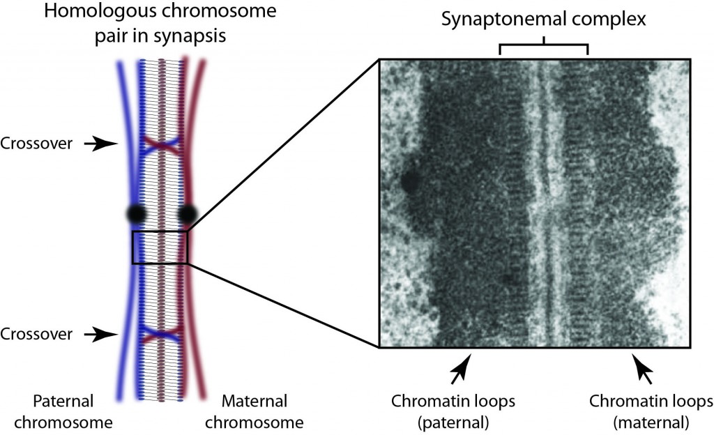
Background: After my PhD, I secured a post-doctoral fellowship from the Royal Commission for the Exhibition of 1851, which I took to the Institute for Stem Cell Research, University of Edinburgh. There I worked with Dr. Sally Lowell studying the molecular triggers of early lineage commitment in embryonic stem cells. It was during this time that I formulated the ideas for my long term research plans for studying the molecular structure and function of the synaptonemal complex in meiosis. Over the last three years, I have initiated this research at the Department of Biochemistry, University of Cambridge, working with Dr. Luca Pellegrini.

Owen has already relocated to Newcastle and has started work in ICaMB


Dr Josana Rodriguez

Research interest: By breaking symmetry, cells are able to generate diversity, migrate, and organise themselves into more complex structures such as tissues and organs. Misregulation of such cell polarity is implicated in a number of human diseases, most notably cancer. Tumour progression is correlated with disruption of epithelial polarity and randomized orientation of the cell division plane caused by misplacement of the mitotic spindle. These observations show the importance of cell polarity for the correct development of an organism and the tight regulation required between cell polarity mechanisms and the cytoskeleton.

My aim is to identify new interactions between cell polarity and the cytoskeleton, and to understand them in a whole organism context during morphogenetic movements and tissue organisation. I would like to extend these studies to analyse the possible implication of these interactions in diseases such as cancer and neurodegenerative disorders.


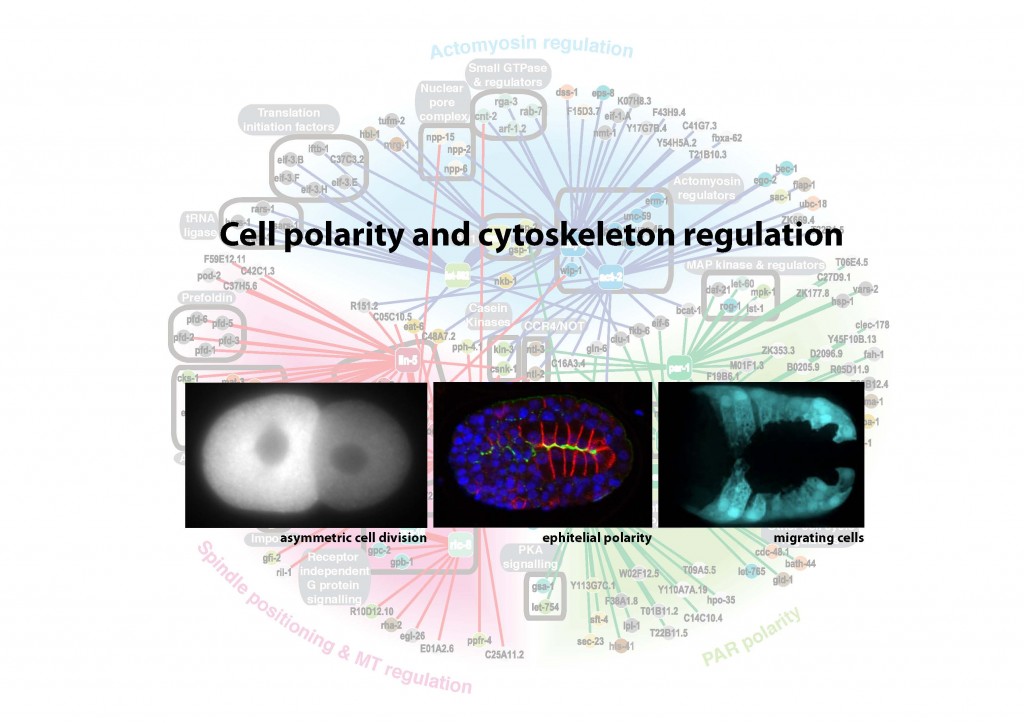
Background: I am currently a postdoctoral research fellow at The Gurdon Institute (Wellcome Trust/Cancer Research, UK) working in the laboratory of Dr. Julie Ahringer (2006 to date). My postdoctoral research identified genes involved in the polarisation of cells through high-throughput genetic screens in C. elegans. I have been a Wolfson College Fellow since 2009 (University of Cambridge).

Josana plans to relocate to Newcastle in July 2014 after completing some ongoing studies in Cambridge


Dr Niall Kenneth

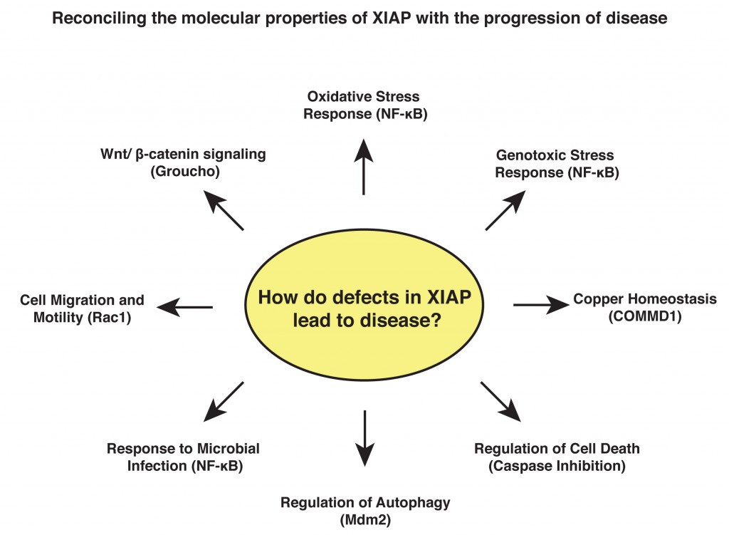
Research Interests:  My work focuses on the signalling properties of a family of intracellular proteins called the IAPs (inhibitor of apoptosis). These proteins were originally characterised as modulators of cell death but have since emerged as key signalling intermediates that regulate a variety of cellular functions. X-linked IAP (XIAP) has been the subject of much recent interest as a possible therapeutic target in cancer due to its greatly elevated expression in tumour cells and its well-documented ability to inhibit cell death. Additional work has identified germline mutations in the XIAP gene that cause a severe primary immunodeficiency known as X-linked lymphoproliferative disorder (XLPD).  My work aims to understand the role played by XIAP in essential cellular processes and to reconcile this with its role in pathogenesis.

Background:  After my PhD, I joined the group of Dr Sonia Rocha at the Centre of Gene Regulation and Expression at the University of Dundee, where I focused on the control of gene transcription following DNA damage and hypoxic stress, regulated by the NF-kB and HIF transcription factors. In 2010, I relocated to the USA to join the laboratory of Professor Colin Duckett, at the University of Michigan, where I have continued to work on transcriptional regulation and developed my interests in the IAP proteins and how they are altered in disease.

Niall will relocate to Newcastle in April 2014


Royal Society University Research Fellowship

The Royal Society URF is one of the most prestigious fellowships awarded to young scientists seeking an independent research career.  We are therefore extremely happy that based on her outstanding postdoctoral work in ICaMB, Yulia Yuzenkova has recently received this award.  

Dr Yulia Yuzenkova

Research Interests: My research is focussed on mechanisms of gene expression in cyanobacteria, one of the most ancient and ecologically important, but under-studied group of organisms on Earth. Approximately 2.3 billion years ago cyanobacteria invented photosynthesis, which transformed all subsequent biological history of Earth. Nowadays they live everywhere where sunlight is available and produce 30% of atmospheric oxygen; furthermore, they can convert inert atmospheric nitrogen to the forms digestible by other organisms. I will be working on transcription in cyanobacteria and its coordination with other major processes in the cell, such as DNA replication and translation.

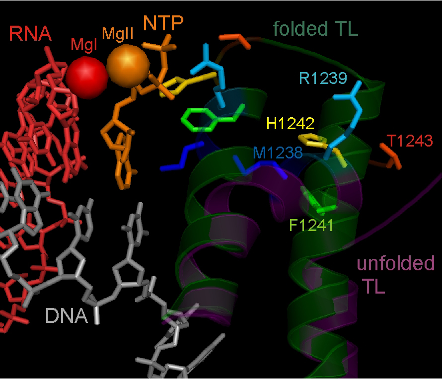
Active centre of the T. thermophilus RNAP elongation complex with unfolded (inactive) and folded (active) Trigger Loop domain conformation.

Background: I did my PhD in the Institute of Molecular Genetics in Moscow and then my first PostDoc in the Waksman Institute in Rutgers, the State University of New Jersey, USA where I performed structure-functional studies of bacterial RNA polymerase.  After moving to Newcastle University, I have been working on a variety of projects investigating the mechanisms of transcriptional regulation by bacterial RNA Polymerase.


New ICaMB Professor

In addition to our new young PIs, we have also recruited a new young(ish) Professor, Jonathan Higgins from the Brigham and Women’s Hospital at Harvard Medical School 

Prof Jonathan Higgins

Research Interests: Cell division is a short but dramatic part of the cell cycle. To ensure precise inheritance of the genetic material, chromosomes must be disentangled, condensed, and then “bi-oriented” on microtubules so that they can be sorted properly into the daughter cells. My lab aims to understand fundamental processes that control these events: specifically, the post-translational modifications of histone proteins that dictate recruitment and displacement of regulatory proteins to and from chromatin during cell division. In particular, my lab has revealed the role of histone kinases such as Haspin in localizing key “error-correcting” proteins to centromeres in mitosis.

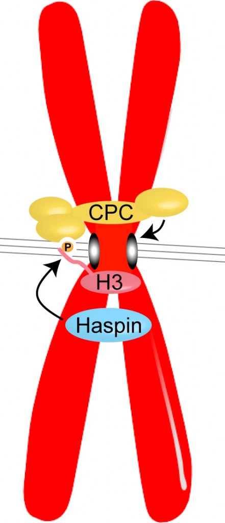
Haspin phosphorylates Histone H3 to create a binding site for the Chromosomal Passenger Complex (CPC) at the centromeres of chromosomes in mitosis. The CPC, which contains the kinase Aurora B, acts to prevent incorrect attachments of microtubules (grey lines) to kinetochores (grey ovals), to ensure the appropriate segregation of chromosomes during cell division.

Background: I was born in Stockton-on-Tees and grew up in North Yorkshire. During my postdoc with Michael Brenner at the Brigham and Women’s Hospital (BWH), Harvard Medical School (HMS), I discovered a novel gene, which turned out to be Haspin, within an intron of the integrin gene I was studying. I started working on Haspin as a side project, and then more seriously when I joined the faculty at BWH/HMS to set up my own research group.

Jonathan will relocate permanently to Newcastle in July 2014

The next generation of ICaMB PIs and Research Fellows

Owen, Josana, Niall, Yulia and Jonathan join a growing group of new PIs in ICaMB, which include 2 further Royal Society URF award winners together with recipients of Wellcome Trust/Royal Society Henry Dale and Career Re-entry Fellowships. We are confident that their talent drive, and enthusiasm will ensure a bright future for research in Cell and Molecular Biosciences in Newcastle.

Dr Suzanne Madgwick: Suzanne is a Wellcome Trust Career Re-entry Fellow researching mechanisms of meiosis

Dr Heath Murray: Heath is a Royal Society University Research Fellow researching the Regulation of Bacterial DNA Replication Initiation

Dr Paula Salgado: Paula is a Lecturer in Macromolecular Crystallography studying the mechanisms of host-pathogen interactions

Dr Claudia Schneider: is a Royal Society University Research Fellow investigating nonsense mediated mRNA decay pathways

Dr Kevin Waldron: is a Wellcome Trust/Royal Society Henry Dale Fellow investigating the role of essential metal ions in pathogenic bacteria


Spills and pills: thrills for a structural biologist

One of the newest recruits to ICaMB is Professor Bert van den Berg, who arrived here in December 2012.  Bert is already off to a great start having been awarded a Royal Society Wolfson Research Merit Award in April.  Here we have asked him to tell us why he decided to join ICaMB and the research that lead up to this prestigious award.

By Bert van den Berg


Bert, looking thrilled

I joined ICaMB in January, coming from the University of Massachusetts Medical School in Worcester, where I was a tenured faculty member in the Program in Molecular Medicine. While I had a great and productive time in this department, after eight years I felt increasingly isolated academically and started to look for another position. ICaMB seemed a great fit for my research interests, with a large number of scientists interested in bacterial biochemistry and cell biology. Since ICaMB was also looking to strengthen its efforts in structural biology, the decision to cross the pond and join ICaMB wasn’t a very hard one. I am happy to be here, and I hope and expect that my expertise in membrane protein structural biology will also be a benefit for the faculty within ICaMB and will lead to successful collaborations.

My lab has been studying protein channels (see below) for about nine years. Determining structures is really the only way to obtain deep insights into protein function. In addition, seeing a new protein structure for the first time is often an “aha!” moment and, at least for me, the closest thing to a true discovery in modern science. In any case, the importance of structural biology for science is clear from the large number of Nobel prizes awarded to the field over the years.

What do the cleanup of oil spills and the treatment of many bacterial infections have in common? The answer is that both processes depend on the efficient passage of bacterial membranes by small molecules.

Oil spills and antibiotics have more in common than you may realise

Gram-negative bacteria are surrounded by two lipid membranes, which are termed plasma membrane and outer membrane. The outer membrane borders the cell and is a very efficient and sturdy barrier that protects the cell from noxious substances in the external environment, such as bile acids in the case of E. coli bacteria living in the gut. However, since bacteria also require nutrients for growth and function, protein channels are present in the outer membrane to allow the uptake of such small molecules. In our work we use X-ray crystallography to determine the atomic 3D structures of the channels, most of which are shaped like hollow barrels. Based on the structures we propose transport models, which we then test by characterisation of mutant proteins.

Many Gram-negative bacteria are able to use industrial pollutants such as oil as food sources, a process called biodegradation. The enzymes that catalyse these remarkable processes are located inside the cell but not much is known about how the pollutants enter the cell in the first place, something that is clearly required before they can be degraded. We study the highly specialised channels that mediate the uptake of these water-insoluble (“hydrophobic”) molecules. In addition, we are interested in discovering cellular adaptations that allow biodegrading bacteria to grow on these toxic compounds. We think that this research may lead to insights that will aid the design of bacterial strains that are optimised not only for bioremediation but also for important other processes such as production of biofuels.

The other main focus of research in my lab is to understand how antibiotics “hijack” outer membrane channels to enter bacteria. Being water-soluble, antibiotics are dependent on protein channels for membrane passage. Bacteria that are under antibiotic pressure will often change or remove the channels through which antibiotics pass, resulting in resistance.

Movie showing ampicillin movement through E coli OmpF protein channel. The view is from the outside of the cell. Movie made by Matteo Ceccarelli (University of Cagliari).

In concert with other mechanisms such as enzymatic degradation and increased efflux by pumps, this acquired antibiotic resistance has the potential to become a huge and global problem in public health. New drugs are therefore urgently needed. The problem is that not nearly enough new drugs are currently in pharmaceutical pipelines, due to the costly and risky nature of antibiotic development. However, pharmaceutical companies are starting to realise that the fundamentals of drug design need to change, and that they have to collaborate with academic labs that are studying the basic biology of small molecule membrane transport.

My lab is participating in an exciting, EU-funded joint venture between big pharma, small biotech firms and academic labs aiming to understand the influx/efflux of drugs in a number of pathogenic Gram-negative bacteria. Beyond the potential benefits for drug design, it is hoped that this project will change the way in which industry and academia work together to benefit public health.


Links

Royal Society Wolfson Merit Awards: http://royalsociety.org/news/2013/new-wolfson-research-merit-awards/

Bert’s ICaMB homepage: http://www.ncl.ac.uk/camb/staff/profile/bert.van-den-berg

Newcastle Structural Biology website: http://sbl.ncl.ac.uk/people/bert_research.shtml

Structural Biologist Nobel Prize Winners: http://www.ebi.ac.uk/pdbe/docs/nobel/nobels.html

IMI TRANSLOCATION project: http://www.imi.europa.eu/content/translocation