Congratulations, Prof Gilbert, FRS!

by Prof. Harry Gilbert, FRS, FMedSci

I was asked to write a blog about my election as a Fellow of the Royal Society. I start by apologising for my ineptitude compared to the “professional” social media people in the Institute. So, what to say? Well, maybe the process might be of interest.

How do you get elected as an FRS?

ICaMB's FRS: Prof Jeff Errington and Prof Harry Gilbert

ICaMB’s FRS: Prof Jeff Errington and Prof Harry Gilbert

You need to be proposed and seconded by current FRS’s. Jeff tried twice to propose me before I went to the USA and a third time (September 2012) upon my return, at which point I said OK. I had to generate a full CV, list 20 papers and include PDF versions of these articles, and the proposers were required to write a three page narrative on my research.

 
Every year material is updated. The first year I wondered what feedback I might get but I soon realized that no one says anything to you. I rapidly put the issue to one side and did not give it any thought.

miebio2014570

‘Wrong Direction’ – Prof Gilbert forms a karaoke boyband with other eminent scientists at a conference in Japan


Looking back on my research it’s evident that most of my science was done with collaborators who are much smarter than me. So, I consider myself to be extremely fortunate to have worked with these people.


The talented people currently working in the Gilbert/Bolam labs

The talented people currently working in the Gilbert/Bolam labs

Bernard_quote

Finally, the election

In late March of this year, I received a letter from the Royal Society marked “In strictest confidence“. I assumed it was yet another reference for someone applying for a grant to the Royal Society and I thought “another job to do”. I read the letter several times and the words “you are on the list of candidates submitted to become an FRS” was hard to digest. When it said you needed 2/3 of the votes to be elected, I became dubious about my chances. On speaking to Jeff, he explained that only one person in the last 150 years has failed to become an FRS at this stage. I was thus confident that not even I could screw this up.

Fun celebrations!

Fun celebrations!

However, keeping it secret for a month before the election took place was extremely hard as I don’t do secrets. I did, however, tell Rosie, my wife, who initially shared my excitement of the news. Her enthusiasm waned somewhat as I kept on about it at home for a day or two. At this point, Rosie said “I hope you don’t become too grand” which ceased any talk about the FRS. I am pleased to say that Rosie’s excitement was rekindled when she realised that she will likely get the opportunity to meet Brian Cox (who also became an FRS this year) who she rates as “very dishy, particularly for a scientist”.


How does it feel to be an FRS?

What are my thoughts about being an FRS? Well, shocked but also excited, although I feel a complete fraud.

Prof Gilbert's speech to his colleagues at ICaMB

Prof Gilbert’s speech to his colleagues at ICaMB

I was very pleased that so many people were able to come and celebrate with me on Friday, I would like to thank you all for coming. I also hope that people will think that “if Harry can become an FRS then the bar is not so high”, and that this will result in other people becoming Fellows in the next few years. In 30 years at Newcastle, I have never worked in a place with so many talented people, and it is clear that ICaMB merits more than two Fellows of the Royal Society.

bobquote2

The ICaMB PhD student symposium: what does it mean to the supervisors?

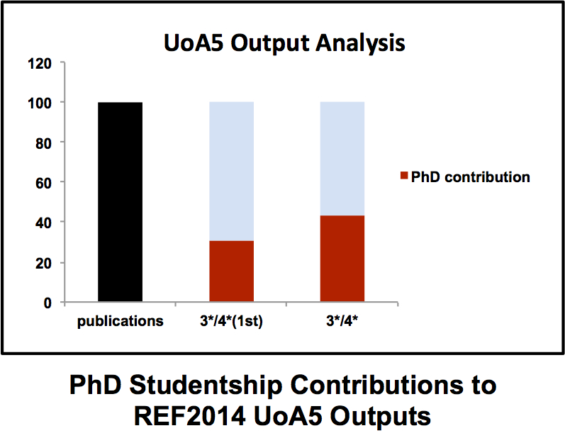
A strong and vibrant PhD programme is essential for any successful academic department. PhD students bring energy and enthusiasm to their projects (well that’s the idea anyway) that frequently reminds many jaded professors contemplating the hell that is ResearchFish why they went into this business in the first place. Typically a PhD student progresses from hesitant first steps in the laboratory to becoming a confident scientist with ownership of their project. Although in the moments before a PhD viva some of that confidence has been known to slip away. The ICaMB Postgraduate Research Symposium is an important showcase that allows our final year PhD students to demonstrate not just the exciting science they have been doing but also just how far they have come over the last 3/4 years. But there is also a serious side to this for ICaMB. In the last REF (where we <cough> did quite well), our PhD students made a major contribution to our returned papers. Brian Morgan has crunched the numbers and discovered that PhD students were first authors on 30% of our 3*/4* submissions (see Figure), including papers in Cell (x2), Nature, Science, Molecular Cell, Nature Chemical Biology (x2) and PNAS (x3). Moreover, in our UO5 return, 3 out of 4 of our Impact cases were underpinned by PhD student research.

Graph

This year, the first day of the symposium was on March 14th with a second to be held on April 29th. We’ve covered the ICaMB PhD symposium before but this year we thought we’d do something different and ask some of the PhD supervisors what this meant to them. All of us are proud of the PhD students that come through our labs, even if, occasionally, there are some grey hair inducing moments on the way. Seeing a final year student confidently discuss their project and answer questions is an important moment for a PhD supervisor. Below we have a varied group of supervisors, from a definitely not jaded professor discussing their final PhD students, to a newer PI discussing their first.

Harry Gilbert: Ana Luis and Jon Briggs

The two final year students from my laboratory who are contributing to the 2016 Postgraduate Research Symposium are Ana Luis and Jon Briggs. These are my last PhD students and it is great to finish with such excellent scientists. Neither student is on the traditional BBSRC/MRC DTP. Jon is supported by the Faculty to work on my Wellcome Trust Senior Investigator Award and Ana is funded mainly from my overheads and more recently by my ERC grant. Jon did a summer placement with Waldemar and his undergrad project in my lab. I was very impressed with Jon and was delighted he was willing to do a PhD with me. I said to Jon his project could be funded by BBSRC, requiring that he did a PIP (Professional Internships for PhD Students) or the Faculty in which case the three month break was not required. Jon said “I want to do science during my PhD and not be distracted by other activities”. Ana wanted to do a PhD with a glycan lab and came highly recommended by one of my previous PhD students, Carlos Fontes from the University of Lisbon, Portugal. So Ana is doing a three year PhD with no MRes, PIPs etc. Some of us, of a certain age, may remember these type of PhDs.

Harry, Jon and Ana
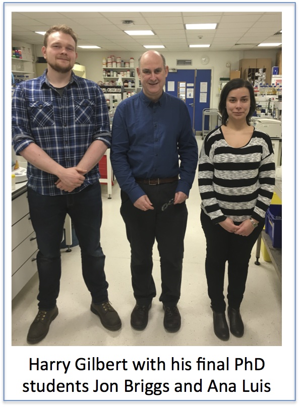
The two students have worked on glycan degradation by the human gut microbial community, or microbiota. Jon is focussed on the biochemistry of selected enzyme systems and the extent to which there is cross feeding of oligosaccharide products generated during the degradative process. Ana, like Jon, has used molecular genetics and biochemistry to explore the enzymology of these glycan degrading processes, while also using X-ray crystallography to study the structure and function of key enzymes.

Both students are remarkable in that they work on their own project within a largish collaboration. This is fine but on a regular basis, well almost weekly, they are given “the opportunity” by their supervisor to alter the objectives of their project almost on a weekly basis. They adapt to these unusual demands brilliantly. We all have tremendous respect for both students; they work extremely hard, are technically excellent, flexible and, most importantly, think carefully about their science, designing and carrying out a series of decisive experiments to resolve critical components of the glycan degrading process. Maybe the best testimony I can provide is that it is not possible to distinguish Ana and Jon from the postdocs in the lab, they are an inspiration to all of us.


Paula Salgado: Adam Crawshaw

Paula and Adam

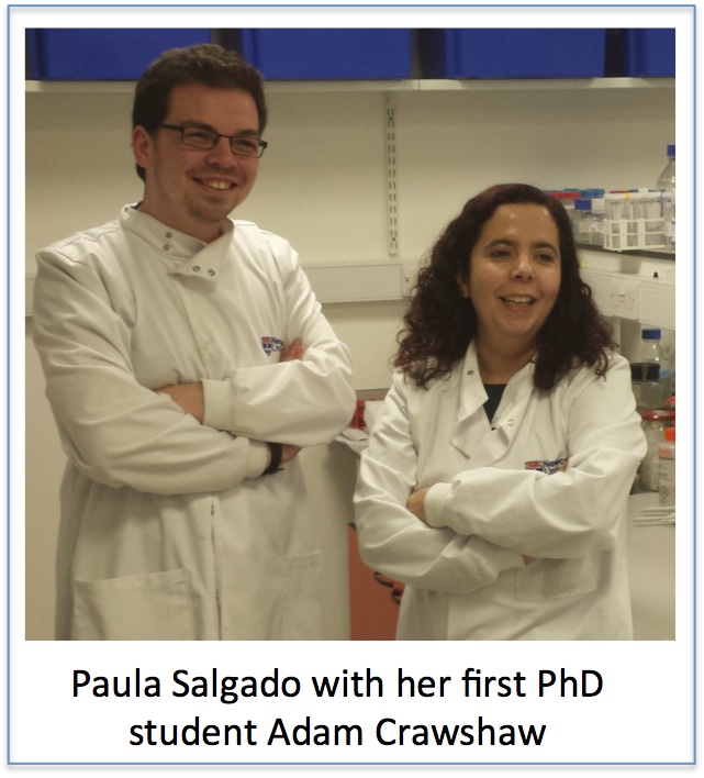
Being your supervisor’s first PhD student can be a mixed bag. You will get a lot of their attention, so help will always be available. But at the same time, as they find their way as independent researchers, any issues will be closer to your own progress than for many of your colleagues. As I saw Adam present his work at the ICaMB Postgraduate Research Symposium, I was reminded of my own journey as a “first student”, several years ago. As I shared my supervisor’s progress, so has Adam shared mine. It has been fantastic seeing him develop, visibly learn and acquire so many skills. Even when his project didn’t go according to plan and experiments were proving hard, he didn’t lose the drive, the enthusiasm. It was with pride that I saw him give a confident talk, answer questions and be humble enough to challenge his own work. He has learned many techniques, from crystallography to circular dichronism, from molecular biology to NMR – he took it all in willingly and enthusiastically. His contribution to understanding several aspects of Clostridium difficile pathogenicity will be in the scientific papers produced, as well as in the future of my “Structural Microbiology” lab. It was great to hear him present all the work he did over the last 3.5 years to our colleagues. Well done!


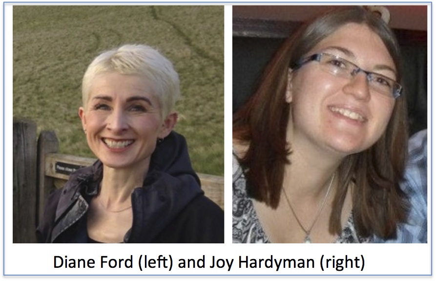
Dianne Ford: Joy Hardyman

 Joy Hardyman’s presentation at the ICaMB Postgraduate Student Research Symposium was on the topic of zinc, which we have studied in our lab for many years. Joy’s PhD research is funded by an MRC studentship, which not only gave Joy the opportunity for research training but also really allowed us to add value to data we collected as part of a BBSRC grant, and generated an additional publication (Hardyman JEJ et al (2016) Metallomics (in press)).

Dianne and Joy

Global zinc
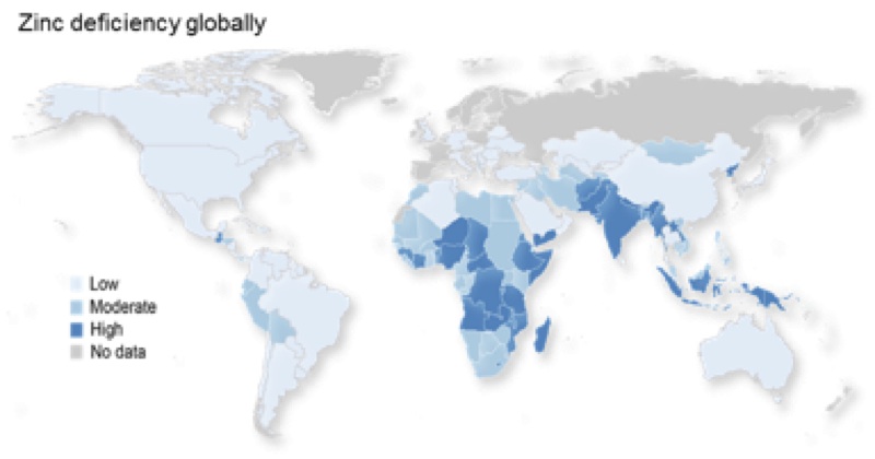
The lab’s focus is the basic cell biology of zinc, which is essential to understanding zinc nutrition. Zinc nutrition is a global health challenge, with an estimated 17.3% of the global population at risk of inadequate zinc intake. Also older people, including here in the UK, are particularly at risk of sub-optimal zinc intake or low plasma zinc concentrations.

Zinc rich foodsAt the end of our BBSRC grant we had some intriguing microarray data that we had set aside because we struggled initially to make sense of it. We had depleted cells of a transcription factor known to have a role in zinc homeostasis (MTF1) then challenged them with zinc, with the aim of identifying the gene targets of this transcription factor. However, rather than see gene responses to zinc being attenuated we saw ‘sensitisation’ of the transcriptome response to zinc. We now know that this is because the usual response of the intracellular protein metallothionein, which effectively ‘mops up’ intracellular zinc, was attenuated because this response is under the control of MTF1.

We all need zincInterestingly, we now think this model, where MTF1 is depleted, may allow us to study what happens to zinc homeostasis in cells as they age, because cells from older individuals have higher levels of metallothionein. Thus, cells with MTF1 depleted may represent a ‘younger’ phenotype. We will now explore the suitability of this as a model of zinc balance in the ageing cell with a view to using it in further research to gain a better understanding of zinc dys-homeostasis in older age.


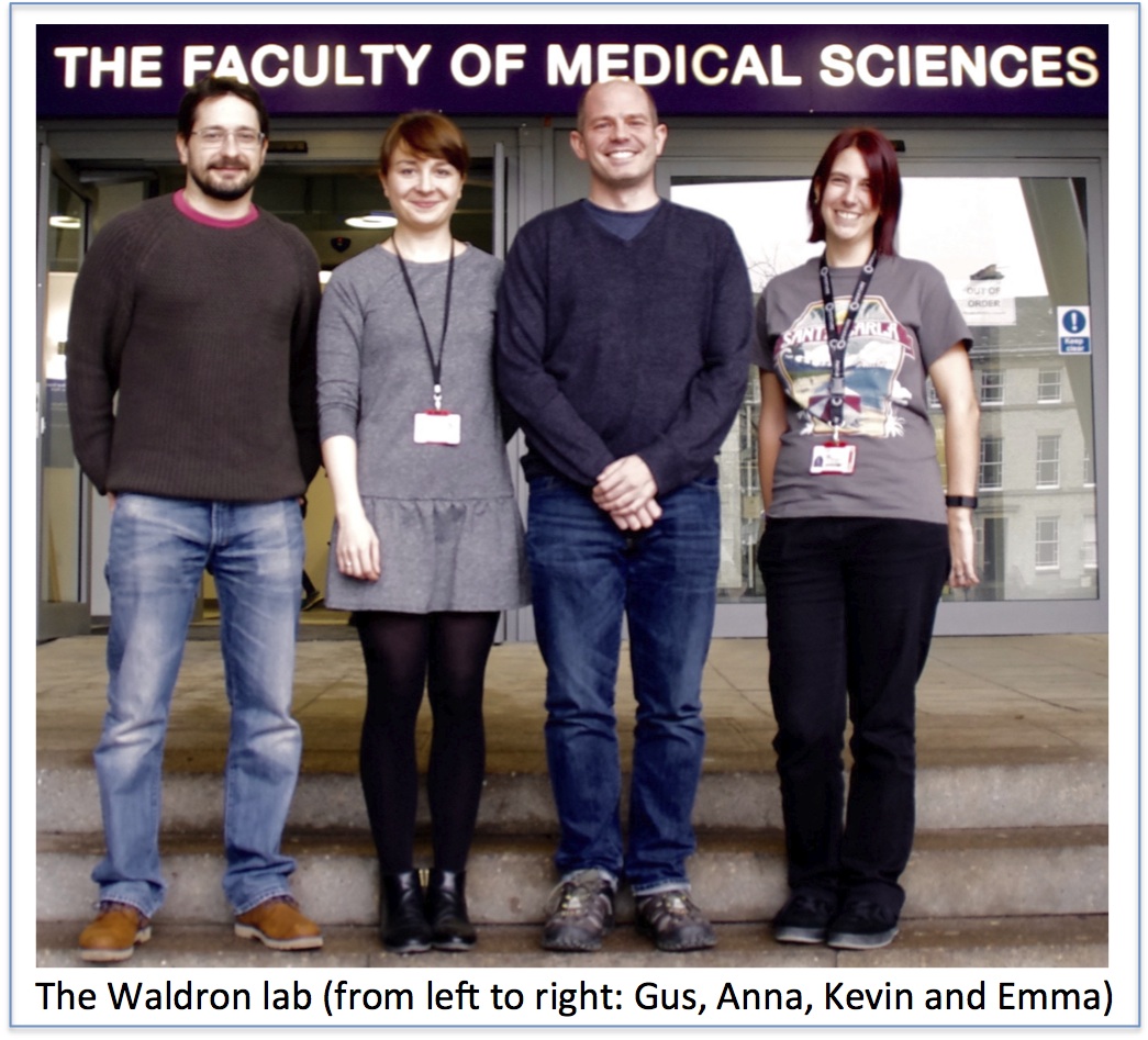
Kevin Waldron: Anna Barwinska-Sendra

Anna’s project began as something of a ‘sideline’ of research in my lab, something that I’d originally initiated when I first started my own independent research back in 2010 but then put on the back burner due to limited resources when my ‘lab group’ consisted solely of me. I re-initiated the project when Anna approached me for a short period of work experience in my lab in 2012. It is a tribute to Anna’s drive and enthusiasm that, within just a few days of her being in my lab, I was keen to keep her on in some form, and I was delighted when she later accepted my offer of a PhD studentship to continue this project in my lab. It’s one of the best decisions I’ve made in my short time as a PI.

Kevin's lab

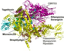
I set Anna the task of determining the metal specificity of the two superoxide dismutase (SOD) enzymes that are encoded within the genome of Staphylococcus aureus. SODs are essential for the bacterial defence against the reactive oxygen species (ROS) superoxide anion, and both of these enzymes were predicted to be manganese-dependent. However it was emerging that, during infection, pathogens such as S. aureus experience host-imposed manganese starvation, a process termed nutritional immunity, which raised the possibility that one or both of these enzymes might be able to use an alternative cofactor for catalysis, most likely iron. Anna has confirmed that one of these enzymes is cambialistic in vitro, which means it is catalytically active with either metal cofactor, something that’s exquisitely rare amongst metalloenzymes. We hypothesise that this cambialistic property of this SOD is a mechanism by which S. aureus is able to circumvent nutritional immunity and resist the onslaught of oxidative attack during manganese starvation.

Anna has been an exceptionally productive student during her time in my lab, and I’ll be sorry to lose her when she completes her studies later this year. She has a bright future in research. Her project also highlights the importance of PhD student projects to a ‘basic research’ lab like mine, as they enable more exploratory, high-risk, high-reward projects such as this, and allow us to take our research in whole new directions that would not be possible within the constraints of traditionally-funded, Research Council grant-based projects.


Jeremy Lakey: Alysia Davies

Alysia poster prize

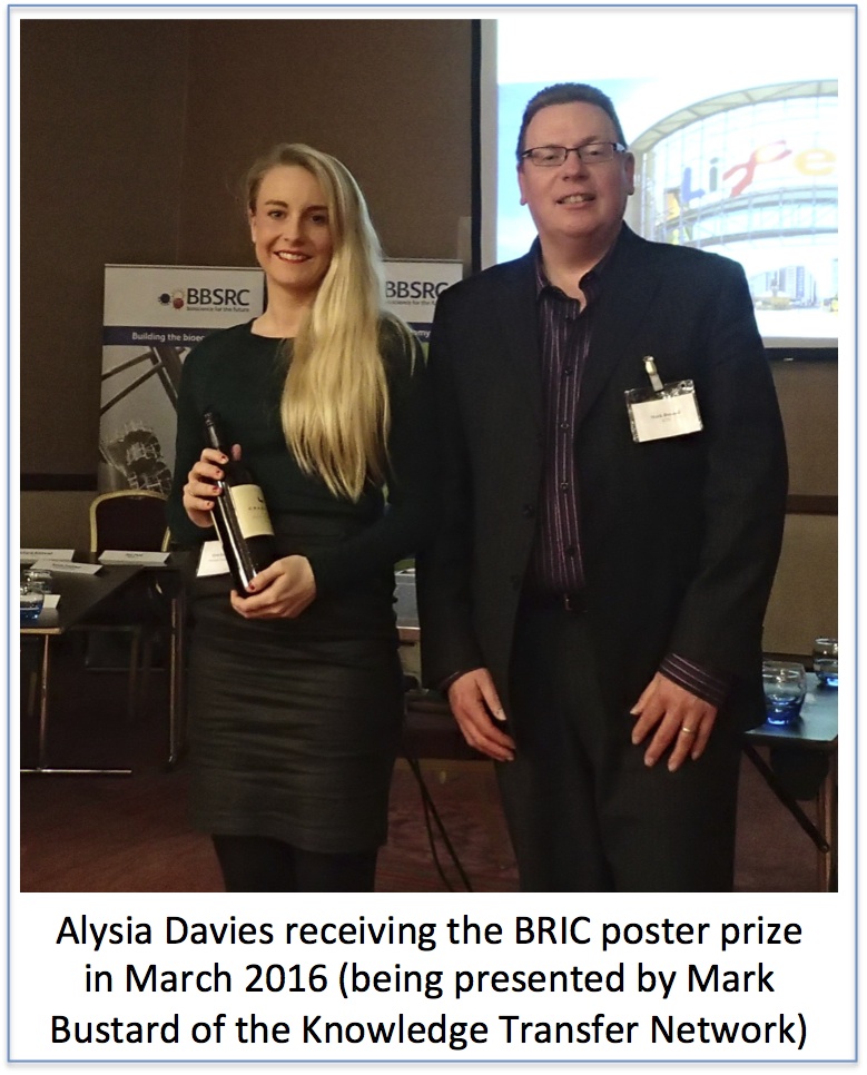
Alysia’s studentship is rather unusual as it is funded by the BBSRC, Bioprocessing Research Industry Club (BRIC) and has a partner company, Pall, who make a huge range of products used in the production and purification of biomolecules. BRIC is a very proactive organisation that meets twice a year for students and post docs to present their work. It also arranges training and skills schools to enhance the employability of the graduates especially those wishing to enter the fast growing bioprocessing industry. This industry is responsible for delivering the next generation of treatments based upon large protein and DNA molecules rather than small molecules such as penicillin. The magic bullets in all this are immunotherapies based upon large proteins called monoclonal antibodies. These are used in the treatment of many conditions such as cancer (Avastin) or rheumatoid arthritis & Crohn’s Disease (Humira). One difference with such large molecules is that they can provoke an immune response which prevents further treatment. Such responses are more common if the proteins stick to each other; a process known as aggregation. Alysia’s project is to develop rapid tests for aggregation so that problem batches can be detected early in the factory and removed. We hope these tests will make the medicines both safer and cheaper.


David Lydall: Joana Rodrigues and Marta Markiewicz

Joana Rodrigues, from Portugal, and Marta Markiewicz, from Poland, will complete their EU (Marie Curie) International Training Network funded PhDs very soon.   On this basis I will vote to stay in Europe.

Lydall lab

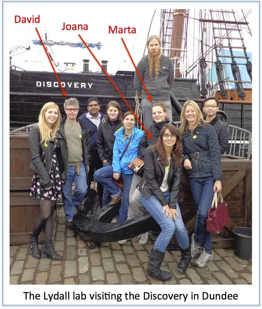
As a supervisor I have been delighted to have Joana and Marta in my lab.  It is really rewarding to have bright, enthusiastic, international members of the lab.  Very often the most rewarding aspect of my job is observing PhD students gain confidence, experience and strength during their comparatively short time in the lab.  I think this has certainly been the case for Joana and Marta and they both gave excellent talks at the symposium.

As is usual, at least in my lab, the projects Marta and Joana have pursued have drifted substantially from where they started.  It is one of the most fun aspects of supervising PhD students that there are few, if any, “milestones” to be met during a PhD.  Despite this chaos, philosophy usually occurs, doctorates are earned and knowledge improves.

Joana and Marta have each worked in budding yeast on proteins that are conserved in human cells and that affect cancer.  Joana has made substantial inroads to our understanding of how the PAF1 complex interacts with and affects telomere function.  Marta has shown how Dna2 protein, known to be important for DNA replication, may play its most important functions at telomeres.  We are just in the process of submitting papers from both Marta and Joana.  They have also each agreed to stay in the lab for a further year to capitalize on all their hard work.

Joana and Marta were recruited to the Codeage International Training Network, which is centred in Cologne (http://itn-codeage.uni-koeln.de).  For all three of us this was our first involvement in such a network.   It has been a lot of fun and we have networked our way from Cologne, to Crete and Milan.  We are looking forward to the final meeting in Crete in September.

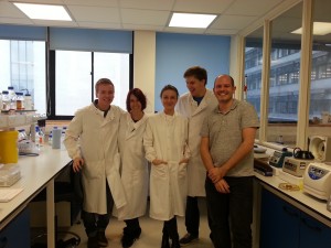
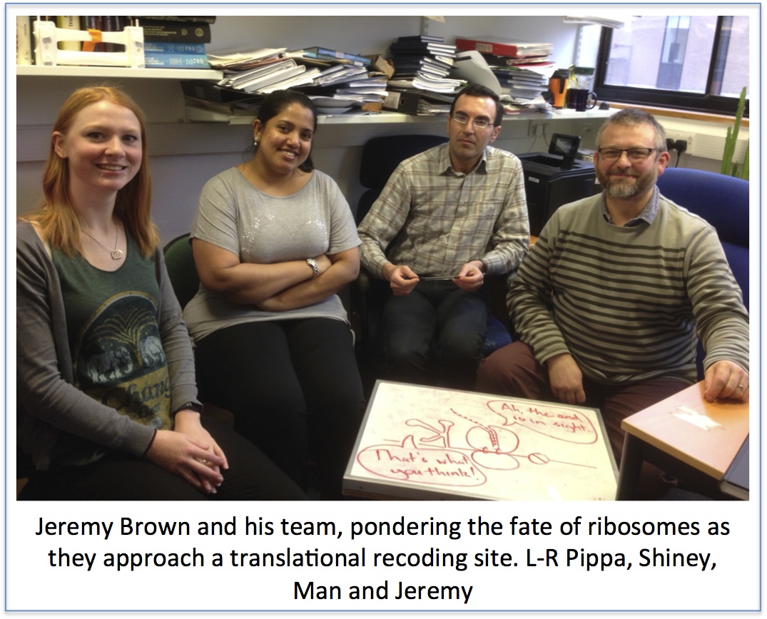
Jeremy Brown: Shiney George and Man Balola

I have 3 PhD students in the final year of their studies. 2 of them, Shiney George and Man Balola gave excellent presentations on their work on translational control of viral gene expression in the Symposium earlier this week. My lab has been dependent on PhD students for the last few years, and I can only thank them for the positive contribution that they have all made to the lab’s research output: without them there would have been little, if any, progress. Nearly all the lab publications in the last few years have had PhD students as first authors, and the current group have generated excellent data for the next papers that we will publish.

Jeremy's lab

My students over the past few years have had very different sources of support, from self-funded through sponsored to Research Council support. This has led me to reflect on the disparities between funding arrangements, and also how this and the structure of PhDs has changed over the years. I was very lucky when I did my PhD – I was the recipient of a Welcome Trust Prize Studentship. These were quite a novelty at that time, a relatively new scheme, with more generous stipend than other studentships, but the same length – 3 years – as most other studentships. At that time pretty much all PhDs were: go to the lab; do the work; learn the trade; write the thesis.

In the years since my time as a PhD student there has certainly been a shift in how PhDs are organised with alterations to funding for students, various add-ons in terms of knowledge and skill training being tried and in some cases discarded, and a move towards longer, 4 year, training. Some of this could be argued to have diluted the important ‘learn the trade’ part of a PhD, though there are clear pros to enhanced training too. Perhaps more worrying though, there is considerable disparity in the provision for students funded from different sources. One obvious issue is the budget for laboratory costs, which is woefully inadequate for many studentships, but much more adequately costed from others. This has to impact at some level the ambition and scope of PhD projects. Another issue is that while the formal length of PhD (i.e. the time from starting to when the book has to be submitted) is pretty much standard at 4 years, there are differences in the length of funding of PhDs. As we know, as staff in ICaMB we can apply for 4 year BBSRC studentships, 3½ year MRC, and faculty studentships (including this year the Research Excellence Academy) that provide 3 years.

There are few level playing fields in life, but as a PhD is an academic qualification one might naively expect that the duration, resources and other support should, where possible, be similar, at least within a country. Why are there such disparities, how confusing must this be for anyone hoping to employ someone with a PhD? Bioscience needs bodies and PhD students are the backbone and key work-force of a good number of laboratories (mine included). There is then strong competition between academics for studentships each year – evidenced particularly by the very large number (>150) of applications for faculty studentships this year – and disappointment for those who are unsuccessful. Conversely there are many aspiring to a career in bioscience for whom a PhD is a key step. So, there are strong reasons to spread the available resource as broadly as possible, and it is easy to rationalise the way in which resources are being used. I would make the comment (my personal view) that at a time when significant efforts being made to even out opportunities at a number of career stages, we should when possible make sure that we do not undermine this at the early stages, by having some PhD students advantaged over others. And this is before the vagaries of supervision, luck and other factors kick in.

Discovery at the Discovery Museum

Aside


Great Hall

Great Hall

Ready to replicate the success of last year’s Away Day, it was en masse outing time for ICaMB again! Time to find out who all the new faces are, and to find out exactly what that person you have a ‘hello and a nod in the corridor’ relationship with actually does at the bench all day. This year we headed to the Great Hall of the Discovery Museum. Despite the leaking roof caused by the downpour outside and the sometimes dodgy acoustics the day was still a success.

Serious faces, this is science

Serious faces, this is science

ICaMB is a fast paced, constantly evolving institute. Everybody is busy with their own research, making a break and a get-together once in a while a vital part of reminding ourselves of the vast range of expertise and diverse set of interests beavering away in our labs and offices. The answer to that tricky problem or that elusive technique is quite possibly just a few yards away.

But also fun!

But also fun!

With that in mind, this year’s Away Day felt particularly important as we welcomed 8 new academic groups to ICaMB from CAV (Campus for Aging and Vitality) as well new IRES fellows and a list of other recent recruits. Drs Victor Korolchuk and Gabi Saretzki from the CAV both spoke at the away day about their interests in neurodegenerative diseases and the role of oxidative stress in the ageing process.

As ever the day was kicked off by the Institute director, Bob (Professor Robert Lightowlers), who gave us a taster of ICaMB’s growth and success stories over the past year. Without breaking into the tune of that well known Christmas song; 7 Vacation Studentships, 6 BBSRC awards, 5 MRC awards, 2 Wolfson awards, 2 Senior Investigator awards and 1 Henry Dale ………. Not to mention all the promotions, outstanding research papers and commercial contracts = 1 happy Bob.

ICaMBAwayDay1(2)

ICaMBAwayDay3


A cell-tastic morning then ensued: the completely dispensable nature of bacterial cell walls (Professor Jeff Errington); the role of NF-kB in the pathogenesis of lymphoma (Dr Jill Hunter); and the cell death independent functions of inhibitors of apoptosis (new IRES recruit Dr Niall Kenneth). The session was wrapped up by Dr Paula Salgado summarising 3.5 years of structural C. difficile research in 15min. Some feat Paula!

Of course just as last year, an absolute highlight of the day were the six, animated, three minute thesis presentations by our brave PhD students ……..  Soon to be followed by the look of horror on several Professorial faces when it was suggested by PAN!C that at next year’s Away Day we have a session of 3 minute PI pitches! We can’t ignore the demands of our PhD students now can we? And congratulations to Mandeep Atwal from the Cowell/Austin lab who against steep competition was awarded the prize for best three minute thesis.

The possibilities of alginate bread?

The possibilities of alginate bread?

A spot of oxidative stress and the evolution of peroxidases by Dr Alison Day, and some lunch completed the morning’s discovery. Though half an hour later and Dr Peter Chater had us all wishing we’d had an alginate packed lunch (and a go with the model gut!). Perhaps the Pearson lab can cater next year’s event? If it’s good enough for the One Show it’s good enough for the ICaMB Away Day.

A major focus of the Away Day is not just to learn about the breadth of exciting research carried out in our institute, but also to learn all about the very latest techniques and expertise ripe for exploitation. This year the focal point of new techniques came from Dr Alex Laude and the Bio-Imaging facility, with some beautiful images and super resolution microscopy techniques, which again left a number of the audience wanting a turn!

P1000375An afternoon transcribing and translating with Dr Danny Castro-Roa; learning about how the crucial nature of cell polarity means we really don’t mix up our arse from our elbow (thank you new IRES recruit Dr Josana Rodriguez); and last but by no means least, how on earth all that DNA manages to faithfully copy and repackage itself time after time from yet another new recruit, Professor Jonathan Higgins.

This completes our diverse and entertaining line-up, just leaving enough time for complementary wine, and the amusement as speakers and audience alike embarrass themselves at the ICaMB quiz (and I hear also in the pub afterwards).

ECRs at ICaMB – Green transcription: how studying Cyanobacteria could change the world

In the latest of our series on the Early Career Researchers of ICaMB we asked Dr Yulia Yuzenkova to tell us about her research and the route that took her from a PhD in Moscow to being awarded a Royal Society University Research Fellowship in Newcastle.

Yulia
Dr Yulia Yuzenkova

My early training was in Moscow, firstly as an undergraduate at Moscow State University and then for a PhD from the Russian Academy of Sciences. However, during my PhD I moved to the USA to study at the Waksman Institute (Rutgers University) with Prof Konstantin Severinov, where I was also a postdoc. My work in the laboratory was dedicated to the molecular mechanisms of inhibition of bacterial transcription by antibacterial peptides and small proteins from viruses. Transcription is the first step and critical regulatory checkpoint of gene expression. In all living organisms transcription is performed by multi-subunit RNA polymerases (RNAP). The central role of transcription in cellular metabolism and the presence of domains specific for bacteria make RNAP an obvious target for antibiotics. Yet, for decades, only one inhibitor of RNAP, rifampicin, has been used in the clinic to treat tuberculosis, while only recently, in 2011, lipiarmycin (fidaxomicin) was approved to eradicate Clostridium difficile. Two is a very small number, but this indicates that there is a good probability of finding new drugs that also work by targetting RNAP. Apart from their clinical significance, antibiotics and inhibitors of transcription in general have been proven to be very efficient molecular tools (see “RNAP details” below for more info).

During my second PostDoc in the lab of Prof Nikolay Zenkin in Newcastle, I have focused on the mechanisms controlling the fidelity of transcription. The copying of genetic information by RNAP is far from being absolutely precise, and RNAP whimsically dislikes reading some DNA sequences, resulting in ‘pauses’. RNAP is able to correct its own mistakes using a proofreading mechanism and the help of small proteins called transcript cleavage factors (explained in detail below). It seems to be important to have at least one cleavage factors; otherwise, the RNAP molecules stop, resulting in “traffic jams” as trailing molecules keep moving and bump into it.

Batch culturing of cyanobacteria

Batch culturing of cyanobacteria

It was therefore a big surprise for me to learn that one large group of bacteria, cyanobacteria (details below), do not encode anything even remotely resembling cleavage factors. I started to look for an explanation for this extraordinary fact. Are there any factors that might compensate for the absence of cleavage factors? Is cyanobacterial RNAP so accurate and at the same time processive that it does not need them? In searching for the answer, I realised that almost nothing is known about the molecular details of transcription in cyanobacteria, and so I decided to apply for a Royal Society University Research Fellowship to try to answer these questions.

Microscopy image of membrane-stained cell of Synechocystis sp 6803

Microscopy image of membrane-stained cell
of Synechocystis sp 6803

In performing preliminary experiments for my application, I became fascinated with cyanobacteria, as they are truly amazing organisms to work with. They are the only prokaryotes that exhibit the classic circadian clock, and are the bacteria with the most complex intracellular organisation. Moreover, they are one of the most ecologically important groups on Earth. They live everywhere where sunlight is available and produce 30% of atmospheric oxygen; some can even convert inert atmospheric nitrogen into a digestible form.


The 2D projection of the membrane structure of cyanobacterium reminds me of a labyrinth

The 2D projection of the membrane structure of cyanobacterium reminds me of a labyrinth

With my Royal Society University Research Fellowship I am planning to investigate the molecular details of the transcription machinery and will look for novel transcription factors that regulate this process. I am also going to test the metal requirements of cyanobacterial transcription, because metal composition of cyanobacterial cells is very different from other bacteria to suit the need of photosynthesis. Another fascinating question is how fast in the cyanobacterial cell, which is tightly packed with photosynthetic organelles, can molecules find their way through the membrane labyrinth.


The nitty-gritty science:

RNAP in detail:

Inhibitors of RNAP as molecular tools for understanding its functions. A wide range of targets of known inhibitors is mapped on the structure of bacterial RNAP. We contributed to understanding the modes of action of inhibitors marked in bold.

Inhibitors of RNAP as molecular tools for understanding its functions. A wide range of targets of known inhibitors is mapped on the structure of bacterial RNAP. We contributed to understanding the modes of action of inhibitors marked in bold.

Studying the antibiotics and inhibitors modes of action have helped us and other groups to discover previously unknown functions and structural domains of RNA polymerase. For example, work on rifampicin shed light on geometry of the RNA exit path, long before the crystal structure of RNAP was solved. Moreover, streptolydigin led to the discovery of the novel catalytic domain, while microcinJ25 confirmed the proposed entry channel for substrates and tagetitoxin provided insight into the mechanisms of RNAP translocation along the template.

Newly synthesised RNA participates in the proofreading  in a ribozyme-like manner. This method of proofreading, probably a remnant from the distant past, is extremely slow. To accelerate proofreading (and to escape from pauses), all 3 domains of life encode non-homologous, but very similarly folded, small proteins called transcript cleavage factors. In E.coli, GreA is an example of a protein that fulfils this role. Some bacteria have several homologs of GreA (in E.coli there are at least 6). It seems to be important to have at least one, because if these cleavage factors are depleted in the cell, the RNAP molecules on the actively transcribed genes stop, obstructing transcription, but also the chromosomal replication machinery moving along the same DNA.

Cyanobacteria:

Cyanobacteria have been hailed as future photobioreactors.  Indeed, when supplied with little more than tap water and light, engineered cyanobacteria can produce all sorts of compounds from sunscreen to biofuels. Cyanobacteria can also be used for environmental applications such as greenhouse gas fixing and cleaning water from industrial pollutants. These initiatives, however, are compromised by slow growth of cyanobacteria and limited knowledge of their basic biology. By putting more effort into research, the potential abilities of cyanobacteria can eventually be harnessed on the industrial level. With this we could make a giant leap towards a future “greener” economy. With a little bit of imagination, it is not hard to envisage cyanobacteria helping humanity to colonise new worlds, and to permit them to inhabit the first lunar and martial greenhouses in not so distant future.

Dr Yulia Yuzenkova’s ICaMB website: http://www.ncl.ac.uk/camb/staff/profile/yulia.yuzenkova

Royal Society University Research Fellowship: https://royalsociety.org/grants/schemes/university-research/

More ICaMB winners! Doctoral Thesis Prize Success.

‘The Faculty of Medical Sciences Doctoral Thesis Prize is a mark of recognition of an outstanding level of achievement by the end of a research doctorate. Prizes are awarded biannually on a very limited basis following nomination by thesis examiners.’ Dr Tim Cheek, Post Graduate Tutor

Doctoral Bling!

Doctoral Bling!

Prizes were first awarded in 2009 and included two ICaMB students, Holly Anderson and Monika Olahova. This was followed in 2011 by David Adams and in 2012 by Graham Scholefield. However, 2013, was an absolute triumph, with three out of only five potential Faculty Prizes being bestowed on theses submitted by ICaMB students. Dr Andrew Foster from Professor Nigel Robinson’s Lab (currently a post-doc in the Robinson lab in Durham), Dr Fiona Cuskin from Professor Harry Gilbert’s lab (currently a post-doc in the Gilbert lab) and Dr Kristoffer Winther from Professor Kenn Gerdes lab (currently a post-doc in Gerdes lab). With their new roles keeping them busy, our 3 winners only just managed to get together recently to be presented with their medals by the Dean of Post Graduate studies. Andrew and Fiona tell us about their past and present research.

Dr Andrew Foster

Dr Andrew Foster

Abstract by Dr Andrew Foster. Achieving metal selectivity is often more difficult than one might first imagine as the inherent chemical properties of metals often mean that a metalloprotein will preferentially select an incorrect metal over a correct one.

My PhD studies involved understanding metal selectivity among a group of proteins called metal sensors. These metal sensing, transcriptional regulators control the expression of genes of metal homeostasis and therefore influence the metallation of other proteins within the cell. I characterised a novel nickel sensor InrS and showed for the first time how metal selectivity could correlate with relative metal affinity across a class of proteins. The nickel sensor InrS has a tighter nickel affinity than the other sensors within the cell, thus InrS responds to nickel activating

Nickel

Nickel

a nickel efflux gene so that the buffered nickel concentration within the cell does not rise high enough to mis-populate the sensors of other metals.

During my PhD studies our lab moved from Newcastle to Durham University but I remained registered at Newcastle. This move was obviously very disruptive but at the same time made me more focussed and determined to make a success of the work in spite of the disruption.

Busy Andrew

Busy Andrew

I am currently working with Professor Nigel Robinson at Durham University. My current work seeks to understand how the affinity of a metal sensor relates to the available concentration of the sensed element within the cell. Our model system involves the nickel sensor I discovered, InrS, and nickel supply to hydrogenase, a nickel enzyme capable of hydrogen production. Metal supply to enzymes will be a key biotechnological challenge as we seek to utilise microbial factories for the production of fuel and other useful products.

Dr Fiona Cuskin.

Dr Fiona Cuskin.

Abstract by Dr Fiona Cuskin. The use of complex carbohydrates in the food industry is wide and varied; a few examples include the use of polysaccharides and oligosaccharides as gelling agents, emulsifiers and fat replacements. Small oligosaccharides are being increasingly used as prebiotics for the vast array of “friendly” bacteria in the gut of both humans and animals. The addition of small fructose oligosaccharides by the food industry into yoghurts, amongst other foods, has been shown to promote a healthy gut flora, which in turn has a positive effect on the host gut health and immune system.

Having been in the lab for just a month my supervisor abandoned me and moved to America. Not to worry I tracked him down and moved there too for a few months. The subject of my PhD was to investigate how bacteria use enzymes called glycoside hydrolases to breakdown complex carbohydrates for utilisation. Part of this was to characterise a glycoside hydrolase that degraded the fructose containing polysaccharide, levan.This glycoside hydrolase contained two

Happy gut!

Happy gut?

modules, the catalytic module and non-catalytic carbohydrate-binding module (CBM). CBMs are usually attached to enzymes that catalyse the breakdown of recalcitrant insoluble substrates to help target the catalytic module to the right carbohydrate. However, the CBM characterised in my PhD bound soluble fructan polysaccharides and potentiated the activity of the catalytic module ~100 fold. This work adds valuable knowledge to how bacteria breakdown complex polysaccharides. This knowledge can be exploited to better inform the use of prebiotics and to also choose enzymes that are efficient for the production of small oligosaccharides from polysaccharides.

We are very proud of our current winners. Who will be in the next batch of Doctoral Thesis Prize winners, adding to a growing list of ICaMB winners?


ECRs at ICaMB: RNA Quality Control


Claudia SchneiderIn the latest of our series focussing on the ECRs in ICaMB, we feature Dr Claudia Schneider. Claudia obtained her PhD from the Philipps-University in Marburg, Germany. She then moved to the UK to work with Prof David Tollervey at the Wellcome Trust Centre for Cell Biology in Edinburgh. In 2011, she was awarded a Royal Society University Research Fellowship and started her own lab at ICaMB. Here, Claudia describes her research, and how being alarmed during her postgraduate studies triggered her long-term research interest.

By Dr Claudia Schneider

Hi, my name is Claudia Schneider, and my Royal Society University Research Fellowship has allowed me to set up my own group here at ICaMB to study enzymes involved in RNA processing and quality control.

I am originally from Germany, where I did my undergraduate studies and my PhD. Many people might think that Germany is the land of cars or lederhosen – but in truth it is really the land of bread (and beer!). It might therefore not come as a surprise that baker’s yeast has become my favourite model organism.

Click on the image to find out how to make these budding buns!

Click on the image to find out how to make these budding buns!

During my undergraduate studies I was first introduced to RNA and I was (and am still) amazed by its many known and still emerging functions in the cell. We now know that almost the entire eukaryotic genome is transcribed, but only a small fraction of the transcripts are protein-coding messenger RNAs (mRNAs). The others are stable and unstable non-coding RNAs (ncRNAs), which are involved in all aspects of gene expression. RNA molecules are often extensively processed before they’re functional, and each processing step is subject to quality control mechanisms. If you want to know more about the life and death of non-coding RNAs, have a look at this recent review.

Yeast has not always been my first choice to study RNA metabolism, since the object of my PhD project in Prof Reinhard Lührmann’s lab turned out to be completely missing in baker’s yeast. Back then I worked on nuclear pre-mRNA splicing, the removal of non-coding introns from precursor mRNAs catalysed by the spliceosome. It was an exciting time in the splicing field: A second low abundance “minor” spliceosome had just been discovered in most multicellular eukaryotes (with the strange and still not readily explainable exception of C. elegans), and this complex is not present in yeast. The minor spliceosome recognises a rare class of introns (<0.5%) with different consensus sequences at the splice sites and has since been linked to a number of human diseases. During my PhD, I purified and biochemically characterised the snRNP components of this unusual pre-mRNA splicing machinery in human and Drosophila cells.

Since then, I have been fascinated by biochemical and enzymatic assays involving RNA such as in vitro splicing assays, where in vitro transcribed pre-mRNAs are mixed with purified spliceosomes. The goal of such an experiment is to observe precise and (hopefully) pretty intron removal in the test tube – but, to my great annoyance, success was every so often hampered by a powerful ribonuclease (RNase) contamination in the assay that completely trashed the precious RNA substrates. Generic and aggressive RNases like RNase A are found on our skin, are incredibly stable and can even survive boiling.

Common decoration on lab surfaces during my PhD

Common decoration on lab surfaces during my PhD

It is therefore fair to say that my scientific career was majorly influenced by constant warnings by my mentor, who told me that all RNases are evil and must be destroyed. However, for my postdoc, I decided to face my fears and look these evil RNases in the eye, in the humble model system yeast. During my time with Prof David Tollervey at the University of Edinburgh, I learned that there are many different types of RNases, and only very few of them are promiscuous and chop RNA to bits.

The majority of RNases are very sophisticated and versatile enzymes. Several RNases are capable of degrading only specific RNA molecules, or only function under certain circumstances, and protein co-factors often assist in substrate recognition. RNases are crucial elements in RNA quality control or surveillance systems, which distinguish aberrant from “normal” RNA molecules. One clinically important RNA surveillance pathway is called “nonsense-mediated decay” or NMD, and this system recognises and degrades a specific class of defective mRNAs to limit the synthesis of truncated and potentially toxic proteins. NMD defects are linked to ~30% of all inherited human diseases (e.g. Duchenne muscular dystrophy and forms of b-thalassemia) as well as certain types of cancer. In addition to quality control/surveillance, where RNAs are mostly completely degraded, a growing number of RNases have been shown to be responsible for precise processing or “trimming” of precursor RNA molecules to produce their functional forms.

Exonucleases were long believed to be the main players involved in RNA recognition and processing/turnover. However, this model was recently challenged by the identification of endonucleases containing PIN (PilT N-terminus) domains, which appear to play key roles in RNA metabolism. Eight PIN domain proteins and therefore putative endonucleases are encoded in the genome of budding yeast, and this includes three largely uncharacterised “orphan” nucleases.

Overall it is still puzzling to me how individual RNases “make the decision” to either completely degrade or carefully process a specific RNA. Given the ever-growing number of non-coding transcripts in the cell, I am also keen to know which RNases are responsible for which substrates and what the so-far uncharacterised putative PIN domain endonucleases in yeast are doing!

To this end, our lab is using an RNA-protein cross-linking method called “CRAC” (UV cross-linking and analysis of cDNA) to identify the targets of PIN domain endonucleases on a transcriptome-wide scale. The CRAC method and the machinery to cross-link yeast cultures were developed by Sander Granneman at the University of Edinburgh, when we were both PostDocs in Prof David Tollervey’s lab. Sander now has his own lab too, and he runs a CRAC-blog. Interestingly, the cross-linking device we are using was originally designed to sterilise sewage water, but it is now also commercially available for research. With this setup, yeast cells are cross-linked while they are growing in culture, which is crucial to identify the often very transient interactions between nucleases and their target RNAs. This system provides a huge advantage over more traditional cross-linkers like the “Stratalinker”, which requires pelleting and cooling the cells on ice before cross-linking. It is, however, also much bigger and takes up a whole bench in the lab – but I guess there is a drawback to everything! In any case: if you want to find RNA targets for your favourite yeast protein – get in touch!!

Cultures of Saccharomyces cerevisiae can be “zapped”, while they are growing: It only takes 100 seconds!

Cultures of Saccharomyces cerevisiae can be “zapped”, while they are growing: It only takes 100 seconds!

Transcriptome-wide RNA-protein interaction analyses generate huge datasets and we use RNA binding and nuclease assays, as well as co-precipitation studies, to validate the in vivo cross-linking results for individual PIN domain endonucleases.

With the help of an ERASMUS exchange student, Franziska Weichmann, who spent 6 months in my lab last year, we have made good progress with two putative PIN domain endonucleases that are linked to ribosome biogenesis. We were able to identify their binding sites on the pre-ribosomal RNAs, as well as co-factors that are important to recruit them into the pre-ribosome. We have also set up an in vitro system with recombinant proteins, and we are currently trying to convince one of them to specifically cleave its proposed rRNA substrate in the test tube – and we are slowly getting there…..

Like the other ICaMB ECRs, who posted on this Blog before, I would like to finish by saying that having my own lab has been an exciting and (on most days) enjoyable adventure so far – and I am looking forward to the next set of challenges…

ECRs at ICaMB: Copying the blueprint of life – Understanding DNA replication


Heath MurrayIn the latest of our series focussing on the Early Career Researchers (ECRs) in ICaMB, we feature Dr Heath Murray.  After completing undergraduate studies at the University of California, Los Angeles and then obtaining his Ph.D. from the University of Wisconsin-Madison, Heath came to the UK to join the lab of Prof Jeff Errington in Oxford. From there he re-located to ICaMB, and in 2009 was awarded a Royal Society University Research Fellowship. Here, Heath describes his research into the mechanisms of DNA replication, and explains why he became interested in this field.

By Dr Heath Murray

Hello, my name is Heath Murray and I’m a Royal Society University Research Fellow in ICaMB’s Centre for Bacterial Cell Biology (CBCB) studying DNA replication. DNA is one of the most important molecules required for life because it encodes the information, or the blueprint, used to build a cell (i.e. the most basic unit of an organism). In order for a cell to create new cells it must synthesize an exact copy of its DNA, an extraordinary process when you consider that the genomes of most cells contain millions of individual DNA subunits!

Bacillus subtilis is a useful model system as it proliferates rapidly and is amenable to genetic, cell biological, biochemical, and structural analyses

Bacillus subtilis is a useful model system as it proliferates rapidly and is amenable to genetic, cell biological, biochemical, and structural analyses

Bacteria are ideal model systems to study this fundamental process because they are much less complex than human cells (e.g. all of their DNA is encoded by a single chromosome, whereas humans have 23), and this allows us to understand how they work at the greatest possible level of detail.

I was introduced to bacteria when I was an undergraduate student and the effect was transformative. My mentors taught me how to add a specific gene (a DNA sequence) to a bacterial cell, and if it worked properly then the bacteria would turn blue!

Bacterial colonies turn blue if they contain a gene that degrades specific sugars.

Bacterial colonies turn blue if they contain a gene that degrades specific sugars.

That basic genetic experiment was one of the coolest things I had ever done, and from that point on I worked hard to learn the trade of “bacterial genetics”.

Today my research group focuses on understanding how DNA replication is controlled so that each new cell will end up containing an exact copy of the genetic material from its predecessor. We employ a wide range of complementary experimental techniques: genetic engineering of bacterial strains, biochemical analysis of purified proteins, and fluorescence microscopy.

In the hot room to check my plates.... I haven't even stopped to take my jacket or backpack off yet!

In the hot room to check my plates…. I haven’t even stopped to take my jacket or backpack off yet!

Fluorescence microscopy is a particular strength of the CBCB because there are several bespoke systems specifically designed for bacterial cells (bacteria are 10-100 times smaller than most human cells). One of the core approaches we use is to genetically engineer a protein we want to study so that it will be fused to a special reporter protein called GFP (Green Fluorescent Protein, originally isolated from jellyfish!) within the cell. Using this approach we can then visualize where our test protein is because it fluoresces when exposed to a specific wavelength of light. Some of our microscopes are so sophisticated that we can observe the location of single proteins and track their movements within living cells.

At the bench.

At the bench.

One of the approaches we often use is to visualize specific regions of the genome within living cells. First, a specific DNA binding protein ( called “LacI”) is genetically fused to GFP. Second, the DNA sequence recognized by LacI (called “lacO”) can be genetically integrated into any location of the genome. Since I study DNA replication, I am particularly interested in the site of the bacterial chromosome where DNA synthesis is initiated (called the “replication origin”). Third, fluorescent dyes are added to cells that bind to the cell membrane and the DNA. Finally, we utilize our fluorescent microscopes to visualize the location of replication origins within individual cells. In the image shown, the live bacterial cells contain chromosomes that are in the process of being replicated, and therefore they have duplicated and separated their replication origins!! This image also emphasizes the fact that although bacteria lack the organelles found in eukaryotic cells, they are nonetheless highly organized (notice how the replication origins are characteristically located at the outer edge of each chromosome).

The GFP protein from jellyfish can be used to fluorescently tag proteins in vivo. Fluorescence microscopy can then be used to localise the tagged protein within the bacterial cell.

The GFP protein from jellyfish can be used to fluorescently tag proteins in vivo. Fluorescence microscopy can then be used to localise the tagged protein within the bacterial cell.

Well that’s it for my first ICaMB blog! I hope you enjoyed hearing about how I became interested in bacterial genetics and about my work on bacterial DNA replication. Please feel welcome to contact me if you have any questions or if you would like further information regarding my research.


Links

Royal Society URF http://royalsociety.org/grants/schemes/university-research/

Centre for Bacterial Cell Biology (CBCB) http://www.ncl.ac.uk/cbcb/


ECRs at ICaMB: Solving 3D puzzles


by Dr Paula Salgado

After nearly one year editing the ICaMBlog, the time has come for me to tell you about my science and work since I joined ICaMB almost 18 months ago.

The fact that it has been 18 months since I moved up North to establish my own research group seems to have snuck up on me… Don’t get me wrong, so much has happened that, if anything, it’s surprising it all took place in 1 and a half years. At the same time, the feeling of a new adventure is still there.

Science is a constant adventure to seek new knowledge, to understand new mechanism, to see new things. In my case, to see into the very core of life’s machines: proteins. I use X-ray protein crystallography to probe the structure of proteins. It’s a bit like solving a puzzle: fitting the pieces of information together until we have a 3D view of the protein.

It is actually fitting that my blog post is the first ICaMB publishes in 2014 as this is the International Year of Crystallography. I could write a lot about it, but for now, I’ll leave you with an amazing video made by the Royal Institution that explains it all – in cartoons! If you want to know more about Crystallography, the Ri has a great collection of videos there, including Prof Stephen Curry’s Friday Evening Discourse, which I strongly recommend.

Freezing protein crystals for data collection at Diamond Light Source. H&S warning: liquid nitrogen is a hazard and we do handle it safely. At this point, I was just dipping the crystals into a small volume, all other procedures handling larger volumes involve wearing appropriate protection.

As a protein crystallographer, I’ve always been interested in proteins that have a relevance to human disease and used this technique to understand their structure and function. In the last few years, I’ve worked on proteins from human pathogens associated with hospital acquired infections, particularly Clostridium difficile and Candida albicans. However, protein structures don’t necessarily give us all the answers and they must be complemented with biochemical studies, as well as in vivo experiments. So my long term goal has become to establish a Structural Microbiology group, where we focus on structure determination of key proteins and complexes involved in pathogenicity as well as on their functional in vivo characterisation.

This is a challenge as it means stepping out of my structural biology comfort zone into the world of microbiology and cell biology. Not that I haven’t stepped out of my comfort zone before – if anything, those are areas that featured strongly during my undergraduate training as a Biochemistry student at the University of Porto in Portugal. In those days, choosing to do protein crystallography as my undergraduate project was the big step into the unknown. A trend that continued as a post-doc, when I joined Dr Cota and Prof Mathews group, a Nuclear Magnetic Resonance (NMR) lab at Imperial College, learning a completely different approach to protein structure determination. And just before coming to ICaMB, I worked in Prof Fairweather’s microbiology lab and always tried to learn a bit about the techniques others were using. So the current idea of bringing structural biology and microbiology expertise together in my lab is the natural evolution of these experiences.

C. difficile cells (green rods) lining the microvilli of the human gut. © Wellcome Trust (CC-BY)

Since joining ICaMB, I’ve focused on 2 main projects, both involving proteins from C. difficile. This spore forming strict anaerobe is resistant to most antibiotics and colonises the gut of individuals whose microbiome has been disturbed by these drugs. It is the most prevalent cause of gastrointestinal infections in hospitals and is a major cause of morbidity and mortality in the hospital environment. Despite recent decreases in the number of deaths and infections as hygiene procedures have improved in the UK, over 1600 people died in England and Wales in 2012 due to C. difficile infections (CDI). It also causes a huge burden to health systems, with an estimated €3,000 million per annum costs in the EU.

C. difficile disease symptoms are caused by the toxins it releases in a process that has been extensively studied over the years. However, the mechanisms of colonisation of the gut and spore formation are poorly understood. So we have been focusing on proteins involved in these two mechanisms.

Firstly, I’ve been trying to determine the structure of SlpA, the main protein constituent in C. difficile S-layer. S-layer is a paracrystalline coat that covers the cell and is presumed to act like a defense mechanism, as well as being involved in gut colonisation. This work, initiated a few years ago in Prof Fairweather’s lab is now a joint collaboration between our two labs and Dr Fagan, at Sheffield University.

SlpA crystals viewed under polarised light (protein crystals are birefringent, unlike salt crystals)


As this protein has tendency to form 2D paracrystalline layers, getting well ordered 3D crystals required for X-ray crystallography has been a challenge, but I have now succeeded in obtaining good crystals. However, other hurdles still need to be overcome to get a structure – but we are getting there!


Our lab: Adam Crawshaw and Paula Salgado

Last year, Adam Crawshaw joined my group as a BBSRC Doctoral Training Programme (DTP) student and we started a new project, looking at a complex between two membrane proteins that are essential for spore formation. As spores are the infectious agents, revealing the molecular details sporulation is important to understand the pathogenicity and infection cycle of C. difficile.

SpoIIQ and SpoIIIAH localise at the membranes of the forming forespore. Green: Membrane; Red: SNAP-tagged proteins.

When spores are first formed, a small cell (forespore) is engulfed by the larger mother cell, physically isolating it from the environment and nutrients in the medium. So, for the forespore to fully mature, it needs a nurturing channel to the mother cell. The two proteins we are studying  – SpoIIQ from the forespore membrane and SpoIIIAH from the mother cell membrane – create this channel. We have already successfully produced recombinant versions of the proteins and shown their interaction in vitro. In collaboration with Prof Henriques at ITQB, Lisbon, we also established their localisation during C. difficile spore formation. Next: crystals! But we are also investigating potential enzymatic activity both in vitro and in vivo, to bring the structure and biology together.

It has been an exciting year and a half – a steep learning curve with many new tasks, from supervising students to managing a lab and teaching undergraduates and postgraduates. The adventure continues, with new challenges and exciting discoveries ahead.


Links

International Year of Crystallography http://www.iycr2014.org/

Royal Institution Crystallography gallery http://richannel.org/celebrating-crystallography

Office for National Statistics (Clostridium difficile data) http://www.ons.gov.uk/ons/rel/subnational-health2/deaths-involving-clostridium-difficile/2012/index.html

European Centre for Disease Intervention and Control on Clostridium difficile  http://www.ecdc.europa.eu/en/healthtopics/healthcare-associated_infections/clostridium_difficile_infection/pages/index.aspx

BBSRC Doctoral Training Programme http://www.ncl.ac.uk/fms/dtp/