How a motile cytoskeleton drives bacterial cell division

seamusIn a recent issue of Science, the discovery of a key mechanism for bacterial cell division was reported. This work was carried out by Dr Seamus Holden’s lab (ICaMB) in collaboration with Professor Cees Dekker (TU Delft), Professor Yves Brun (Indiana University), Professor Mike VanNieuwenhze (Indiana University) and Professor Ethan Garner (Harvard University). Here, Seamus tells us about this discovery and what its implications could be for antimicrobial research.

Bacterial cell division is a lovely mechanistic problem in biology: how do the simplest living organisms build a crosswall at mid cell, against very high outwards pressure (think of a racing bike tyre), without bursting?  A ring of protein filaments forms around the future division site, and enzymes associated with this ring build a new crosswall that cleaves the bacteria in half. But what has remained completely mysterious is how these proteins work together as a single nanoscale machine to cut the bacterial balloon skin (cell wall) in two.

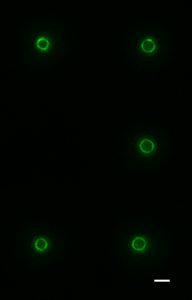
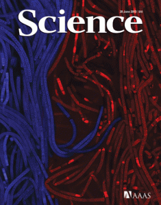
Cytoskeletal proteins FtsZ in live bacteria imaged in vertical nanocages

Cytoskeletal proteins FtsZ in live bacteria imaged in vertical nanocages

Working together with collaborators in Delft, Indiana and Harvard, we tracked the organization and motion of key division proteins as they build the dividing crosswall, and the organization of the newly built crosswall itself. We began by examining the motion of FtsZ, a cytoskeletal filament that is required for cell division – cytokinesis – in bacteria and is related to the tubulin cytoskeletal protein found in eukaryotic cells. Using high-resolution microscopy techniques, we found that FtsZ filaments move around the division site, traveling around the division ring. We imaged the motion of individual cell wall synthesis enzymes, and saw that the synthesis enzymes ride on FtsZ filaments, building new cell wall as they travel along the division site. This causes the cell wall to be synthesized in discrete sites that travel around the division site during cytokinesis, a process which we were able to observe directly by using dyes that label the bacterial cell wall. Using a variety of experimental techniques, we were able to speed up or slow down how fast FtsZ rotated around the cell. Strikingly, we found that the speed of FtsZ filament motion determines how fast the cell can divide. When FtsZ moves more rapidly, cell wall is produced more quickly, and cytokinesis happens faster. This shows that the motion of FtsZ is the critical overall controller of cell division.

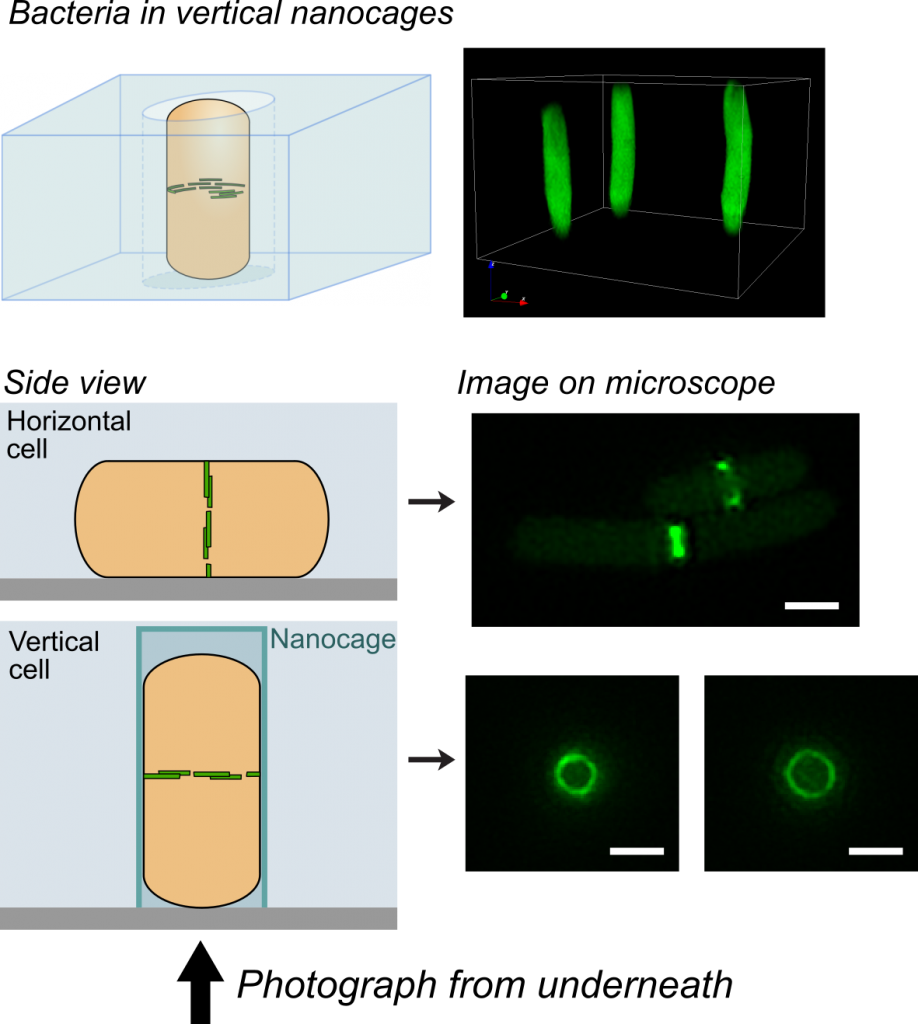
One challenge that we faced was trying to look at the division proteins in actively dividing cells. At the earliest stages of division, it was possible to image division protein organization because the proteins in the partially assembled ring are sparsely distributed. However, a new strategy was required to measure how the dense protein network of actively dividing cells was organized. Normally, bacteria are immobilized flat on a microscope slide, and imaged from underneath, but unfortunately this places the division ring side-on, obscuring the motion and organization of division proteins. To solve this problem, we used nanofabrication technology, originally developed to manufacture computer chips, to create tiny gel nanocages to trap bacteria in an upright position.

Bacteria trapped in vertical nanocages

Bacteria trapped in vertical nanocages

By trapping individual bacteria upright, we were able to rotate the cell division ring so that it was fully visible on our high resolution microscope. This revealed the dynamic motion of FtsZ filaments as they travel around the entire division site:

Together, these results revealed the basic mechanistic principles of bacterial cell division: that the building of the division crosswall is orchestrated by moving cytoskeletal filaments.  Previously, the cytoskeleton was thought to serve as a static scaffold, recruiting other molecules and perhaps exerting some force to divide the cell. This new work demonstrates that all the components of cell division are in constant, controlled motion around the division site, driven by the fundamental dynamics of the cytoskeleton.

In the longer term, this study could open up novel antibiotic targets. Based on the discovery that the treadmilling motion of the bacterial cytoskeleton is critical for division, it may be possible to develop new drugs that specifically inhibit this motion, similar to how the chemotherapy drug taxol suppresses the motion of the cytoskeleton in cancer cells.

==================================

Links

Explanatory animation: https://youtu.be/6dq2_gqKPfU (Animation credit TU Delft / Scixel)

Nanocage Video: nanocage-movie-2.

Science report: http://science.sciencemag.org/content/355/6326/739

Press release: http://bit.ly/2kwsnJf

Origins of life in Newcastle

ICaMB’s Prof Jeff Errington organised and hosted an impromptu symposium on the origins of life at the Centre for Bacterial Cell Biology (CBCB) on Wednesday 18th January 2017. About 80 people attended, hearing 11 talks from a mixture of Newcastle and international speakers, including a number of guests who had travelled over from Japan for the meeting. The program was arranged more or less in “chronological” order, starting with the origins of the solar system 4-6 billion years ago, and ending (still almost 2 billion years in the past) with the emergence of the eukaryotes. The meeting sparked several very lively discussions, perhaps reflecting the difficulty of doing standard hypothetico-deductive experiments on the topic, in the absence of time travel technology! Nevertheless, the day was a great success and is likely to lead to new international collaborations and funding opportunities.

By Jeff Errington

My emerging interest in the subject has two origins. First, through ageing and trying to find a reason for existence before existence disappears! Second, the lab’s work on L-form bacteria (see Box), which has attracted much interest from the origins of life scientific community.

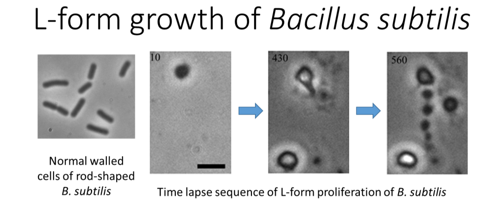
L-form bacteria use a seemingly primitve mechanism of replication.

L-form bacteria use a seemingly primitive mechanism of replication. L-forms are cell wall deficient bacteria, which turn out to replicate by a slightly bizarre, seemingly haphazard mechanism involving membrane blebbing and tubulation. The process provides a model for how primitive life may have proliferated billions of years ago, before the invention of the cell wall.


The latest findings have led to a number of fascinating new scientific contacts, and about a year ago, Prof Shige Maruyama, who heads a major Japanese research institute dedicated to origins of life work called the Earth-Life Science Institute (ELSI), made contact, proposing discussion around possible collaborations. After a series of small meetings in Newcastle and my visit to Tokyo, momentum began to emerge, culminating with the proposal for a major workshop in Newcastle, with half a dozen or so ELSI members planning to attend.

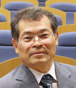
Prof Shige Maruyama, ELSI, Tokyo, Japan

Prof Shige Maruyama, ELSI, Tokyo, Japan

As discussions developed, I identified various experts in Newcastle with complementary expertise and interests in the general area, and the idea for a full blown symposium took shape. There was even time to identify a top class international “guest” speaker, Prof Bill Martin from Dusseldorf, who came over at short notice to give the concluding talk.

This is not the place to go through each talk in detail. However, from my perspective, what I hope people took away from the meeting would have included the following general points.

First, the problem is amazingly multidisciplinary, with important contributions from astrophysicists, geochemists, organic chemists, microbiologists (structure/function, metabolism and physiology) and evolutionary bioinformaticians. Second, we still have a very hazy understanding of many of the early events in the earth’s planetary history, e.g. when did the water arrive and how much? Third, it is clear that microbes were responsible for huge changes in planetary chemistry, particularly oxygenation but also that planetary composition must have reciprocally influenced microbial evolution.

Prof Bill Martin, Dusseldorf, Germany

Prof Bill Martin, Dusseldorf, Germany

The day concluded with a very nice dinner at the Jesmond Dene House Hotel, supported by Newcastle University and hosted by Pro-Vice Chancellor Prof Nick Wright. I’m sure that the original owner of the house, Lord Armstrong, would have approved of the day (for example, I gather that his company won the contract to build ships for the Japanese Navy 120 or so years ago). I’m also sure that as a Fellow of the Royal Society (elected in 1843) he would have been acquainted with Charles Darwin and perhaps they too had interesting conversations about the origins of life in their own time frame.

Another Cell-ebration

Heath MurrayKevin WaldronLast year we brought you details of the inaugural CBCB Symposium. In July the second CBCB symposium was held, and today we hear from the organisers, Kevin Waldron and Heath Murray, about this latest successful event.
The idea for an annual Centre for Bacterial Cell Biology (CBCB) symposium was originally conceived in 2013, and aimed to showcase both the high quality and the immense breadth of research activity that goes on in this unique Centre. It would also be an excellent opportunity to bring together the CBCB research community based in both the Medical School and in the Baddiley-Clark building to discuss their work and build future collaborations.

It wasn’t long after our 2014 Symposium was over when the organising team (Bernie Shaw, Heath Murray, Kevin Waldron and Jeff Errington) began planning for this year’s second event. Obviously we were delighted with the success of that first meeting, but of course it also applied a little pressure on us as organisers; this year’s event had to achieve a similar level of success. Fortunately, the feedback we had received from the 2014 event included a number of constructive suggestions from the CBCB community about how we might be able to improve the Symposium, and we tried to incorporate as many of these ideas as possible.

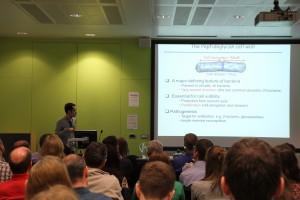
One of our postdoctoral researcher speakers, Yoshi Kawai, addresses the Symposium audience on the subject of L-forms

One of our postdoctoral researcher speakers, Yoshi Kawai, addresses the Symposium audience on the subject of L-forms

One suggestion was to include a number of more junior speakers in the Symposium Programme as well as PIs, and we are grateful to those postdocs who volunteered to present their work to the CBCB audience. Alexander Egan told us about his research in the Vollmer lab on the proteins that coordinate biosynthesis of the cell envelope during growth and division. Yoshi Kawai of the Errington lab explained  how L-forms, bacteria that lack their cell wall, can be produced in the lab and how they propagate in a manner independent from the known bacterial cell division machinery, as well as speculating on their implications for early life forms on Earth. Marcin Dembek of the Salgado lab contrasted the mechanisms that govern sporulation in Clostridium difficile, a pathogen that primarily causes infections via spores, and the model organism Bacillus subtilis. Finally Didier Ndeh described his research in the Gilbert lab on how gut bacteria degrade the most structurally complex dietary polysaccharide known, rhamnogalacturonan II. PI speakers covered further topics relating to antibiotic discovery and their mechanisms of action and synthetic biology.

In addition to our CBCB researchers, we also again invited two high-profile external speakers. The day started with Mark Leake (University of York) who told us about his research using state-of-the-art microscopy for in vivo imaging of single molecules within the bacterial cell. And the Symposium was concluded by John Helmann (Cornell University) on the subject of transcriptional stress responses in one of CBCB researchers’ favourite model organisms, Bacillus subtilis.

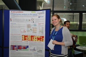
Poster prize winner Lauren Drage

Poster prize winner Lauren Drage

Another of the suggestions that we incorporated into the Symposium schedule this year was a poster session, which was accompanied by light refreshments (of course!) immediately after the day’s talks. We had a great turnout, with more than 20 posters on display, and the session generated a lot of lively scientific discussion. Again the Symposium organisers are very grateful to all those members of CBCB who participated in the poster session. We awarded three poster prizes, with congratulations to winner Lauren Drage for her excellent poster describing her research in the Aldridge lab looking for biomarkers for diagnosis of urinary tract infections, and to our two runners-up, Martin Sim (Wipat lab) and Clare Wilson (Errington lab); and of course thanks to our poster judges, Lucy Eland and Yulia Yuzenkova.

Finally, we all got to enjoy an informal barbecue dinner and drinks, where the science discussions could continue into the evening.

Jeff Errington and John Helmann in post-symposium discussions

Jeff Errington and John Helmann in post-symposium discussions

Planning has already begun for next year’s Symposium, which will be held on the 8th July 2016, and will feature two more external keynote speakers, Christine Jacobs-Wagner (Microbial Sciences Institute, Yale University) and Prof. Tracy Palmer (Molecular Microbiology, University of Dundee). We welcome your feedback too, so if you attended this year’s Symposium and you have any suggestions about how we might improve next year, please let us know.

A CBCB Cell-ebration

Heath MurrayKevin WaldronEarlier this month, the Centre for Bacterial Cell Biology held its inaugural Symposium. Here, the CBCB’s Heath Murray and Kevin Waldron tell us about what happened at the event.

One of the aspects of ICaMB that makes it a unique institute is the Centre for Bacterial Cell Biology (CBCB), a group of researchers who are focused on understanding fundamental biological questions using bacteria as model organisms. The CBCB was founded by Professor Jeff Errington FRS and is the world’s first major research centre with a focus on bacterial cell biology. Since its inception, CBCB has relocated to a purpose-built £30 million facility in the Baddiley-Clark Building, and has grown to include more than 20 different research groups. In a relatively short time, CBCB members have made outstanding contributions to our understanding of numerous aspects of fundamental cellular processes in a wide range of bacteria.

Prof Kenn Gerdes from the CBCB discusses how bacteria can form dormant variants that evade the immune defence response.

Prof Kenn Gerdes from the CBCB discusses how bacteria can form dormant variants that evade the immune defence response.

In order to recognise the success and the breadth of science being generated in the Centre, we recently held the inaugural CBCB Symposium on July 9-10. More than 120 members of the CBCB community participated in the two-day event, underscoring the critical mass of researchers at Newcastle University working within the field. This excellent turnout certainly contributed to the overall success of the event.

Research themes covered by talks from group leaders in the CBCB included sporulation, infection, persistence, biofilms, metabolism, motility, and morphogenesis. We also heard about the emerging subject of synthetic biology, where bacterial organisms will be programmed much like computers to perform discrete biological tasks.The CBCB Symposium was highlighted by inspirational talks from three distinguished external scientists, Jan Löwe (Laboratory of Molecular Biology, Cambridge), Mervyn Bibb (John Innes Centre, Norwich), and Simon Foster (Department of Molecular Biology and Biotechnology, Sheffield).

Prof Simon Foster explains how the superbug Staphylococcus aureus grows and divides.

Prof Simon Foster explains how the superbug Staphylococcus aureus grows and divides.

Professor Löwe discussed his work using a range of biochemical and structural approaches to analyse the bacterial cell division and morphogenesis machinery. Professor Bibb explained how his lab utilises a combination of next generation DNA sequencing and bioinformatics with classical genetic analysis to discover novel antibiotics. Professor Foster showed how studies on the fundamental aspects of bacterial cell biology can be harnessed to better understand host-pathogen interactions that can eventually be translated into vaccine development, with his focus on the ‘super bug’ Staphylococcus aureus..

Participants hold discussions over dinner and drinks following the Symposium.

Participants hold discussions over dinner and drinks following the Symposium.

At the end of the Symposium participants gathered together for dinner and drinks in the informal setting of the Forum. This provided an interactive end to the event that allowed researchers throughout the CBCB to meet one another, discuss the amazing science, and develop connections.

ECRs at ICaMB: Copying the blueprint of life – Understanding DNA replication


Heath MurrayIn the latest of our series focussing on the Early Career Researchers (ECRs) in ICaMB, we feature Dr Heath Murray.  After completing undergraduate studies at the University of California, Los Angeles and then obtaining his Ph.D. from the University of Wisconsin-Madison, Heath came to the UK to join the lab of Prof Jeff Errington in Oxford. From there he re-located to ICaMB, and in 2009 was awarded a Royal Society University Research Fellowship. Here, Heath describes his research into the mechanisms of DNA replication, and explains why he became interested in this field.

By Dr Heath Murray

Hello, my name is Heath Murray and I’m a Royal Society University Research Fellow in ICaMB’s Centre for Bacterial Cell Biology (CBCB) studying DNA replication. DNA is one of the most important molecules required for life because it encodes the information, or the blueprint, used to build a cell (i.e. the most basic unit of an organism). In order for a cell to create new cells it must synthesize an exact copy of its DNA, an extraordinary process when you consider that the genomes of most cells contain millions of individual DNA subunits!

Bacillus subtilis is a useful model system as it proliferates rapidly and is amenable to genetic, cell biological, biochemical, and structural analyses

Bacillus subtilis is a useful model system as it proliferates rapidly and is amenable to genetic, cell biological, biochemical, and structural analyses

Bacteria are ideal model systems to study this fundamental process because they are much less complex than human cells (e.g. all of their DNA is encoded by a single chromosome, whereas humans have 23), and this allows us to understand how they work at the greatest possible level of detail.

I was introduced to bacteria when I was an undergraduate student and the effect was transformative. My mentors taught me how to add a specific gene (a DNA sequence) to a bacterial cell, and if it worked properly then the bacteria would turn blue!

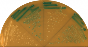
Bacterial colonies turn blue if they contain a gene that degrades specific sugars.

Bacterial colonies turn blue if they contain a gene that degrades specific sugars.

That basic genetic experiment was one of the coolest things I had ever done, and from that point on I worked hard to learn the trade of “bacterial genetics”.

Today my research group focuses on understanding how DNA replication is controlled so that each new cell will end up containing an exact copy of the genetic material from its predecessor. We employ a wide range of complementary experimental techniques: genetic engineering of bacterial strains, biochemical analysis of purified proteins, and fluorescence microscopy.

In the hot room to check my plates.... I haven't even stopped to take my jacket or backpack off yet!

In the hot room to check my plates…. I haven’t even stopped to take my jacket or backpack off yet!

Fluorescence microscopy is a particular strength of the CBCB because there are several bespoke systems specifically designed for bacterial cells (bacteria are 10-100 times smaller than most human cells). One of the core approaches we use is to genetically engineer a protein we want to study so that it will be fused to a special reporter protein called GFP (Green Fluorescent Protein, originally isolated from jellyfish!) within the cell. Using this approach we can then visualize where our test protein is because it fluoresces when exposed to a specific wavelength of light. Some of our microscopes are so sophisticated that we can observe the location of single proteins and track their movements within living cells.

At the bench.

At the bench.

One of the approaches we often use is to visualize specific regions of the genome within living cells. First, a specific DNA binding protein ( called “LacI”) is genetically fused to GFP. Second, the DNA sequence recognized by LacI (called “lacO”) can be genetically integrated into any location of the genome. Since I study DNA replication, I am particularly interested in the site of the bacterial chromosome where DNA synthesis is initiated (called the “replication origin”). Third, fluorescent dyes are added to cells that bind to the cell membrane and the DNA. Finally, we utilize our fluorescent microscopes to visualize the location of replication origins within individual cells. In the image shown, the live bacterial cells contain chromosomes that are in the process of being replicated, and therefore they have duplicated and separated their replication origins!! This image also emphasizes the fact that although bacteria lack the organelles found in eukaryotic cells, they are nonetheless highly organized (notice how the replication origins are characteristically located at the outer edge of each chromosome).

The GFP protein from jellyfish can be used to fluorescently tag proteins in vivo. Fluorescence microscopy can then be used to localise the tagged protein within the bacterial cell.

The GFP protein from jellyfish can be used to fluorescently tag proteins in vivo. Fluorescence microscopy can then be used to localise the tagged protein within the bacterial cell.

Well that’s it for my first ICaMB blog! I hope you enjoyed hearing about how I became interested in bacterial genetics and about my work on bacterial DNA replication. Please feel welcome to contact me if you have any questions or if you would like further information regarding my research.


Links

Royal Society URF http://royalsociety.org/grants/schemes/university-research/

Centre for Bacterial Cell Biology (CBCB) http://www.ncl.ac.uk/cbcb/


Science Minister visits Centre for Bacterial Cell Biology


by Dr Heath Murray 

On June 27 the RH David Willetts MP, Minister for Universities and Science, visited the Centre for Bacterial Cell Biology (CBCB) to hear about how research on bacteria can lead to: development of novel antibiotics, design of synthetic biological systems, and even understanding the origins of life on earth. Dr Heath Murray (CBCB & ICaMB) tells us more about this visit.

Mr. Willetts was given a guided tour of the new Baddiley-Clark building by the director of the CBCB, Prof Jeff Errington.

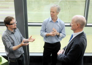
Jeff (left) outlines CBCB research to David Willetts (right), with Heath (middle back) paying close attention

Jeff discussed why he left Oxford University after 25 years to start the CBCB at Newcastle, the first Centre of its kind in the UK to provide a world-class facility in which to carry out fundamental research on bacterial cells. During the tour Jeff highlighted how the localised network of international researchers at the CBCB, working on biological problems in model bacterial organisms provides an unparalleled setting in which to exchange ideas and to benefit from related advances in microbial cell biology. While walking around Jeff noted how the open plan of the Baddiley-Clark building promoted interactions amongst the various research groups, thereby creating a uniquely stimulating environment for the scientists that work there.

This was a very fruitful visit with interesting discussions, as highlighted by Jeff: “I was impressed at how quickly the Minister picked up the key biological points we wanted to make, such as about how our work impacts on thinking about the origins of life!

An image similar to those seen by David Willetts showing severe DNA segregation defect in a mutant Bacillus subtilis strain, observed using epifluorescent microscopy. (DNA: blue; origin of replication: green, cell membrane: red)


I then demonstrated the bespoke microscopes available within the CBCB to the Minister, highlighting how the small size of bacterial cells (only a few micrometers) makes microscopic analysis technically challenging and how the CBCB is utilizing state-of-the-art super-resolution microscopes to overcome this difficulty. I also explained how researchers use genetic engineering to fuse their “proteins of interest” to the Green Fluorescent Protein (GFP) from the jellyfish Aequorea victoria, thus creating tools to visualize the localization of proteins or nucleic acids within living bacterial cells using fluorescence microscopy.


Heath explains the potential applications of the research to the Science Minister


The Minister was keen to see the live demonstration of our fluorescent microscope and seemed amazed by how clearly the organization of the bacterial chromosomes was immediately apparent. He quickly appreciated that interfering with this process might have application in the development of new antibiotics.


We were all left with the clear feeling that Mr. Willetts enjoyed hearing about the science taking place within the CBCB and how this fundamental research provides insights crucial for the discovery and development of new antibiotics, as well as providing solutions to a wide range of industrial and environmental problems. “It was an interesting meeting – very reassuring to hear that the Minister is keen to make sure that Government continues to invest in Blue-Skies Research”, Jeff concluded.


ICaMB Research Update: Zenkin lab Science paper

Congratulations to ICaMB and CBCB researchers Soren Nielsen, Yulia Yezenkova and Nikolay Zenkin and  who have a paper published in the prestigious journal Science today.

Nikolay Zenkin: RNA Polymerase researcher

There are three RNA Polymerases in Eukaryotic cells and although much attention focuses on the role of RNA Polymerase II, since it transcribes mRNAs from protein encoding genes, it is easy to forget that the majority of transcription in the cell is accomplished by RNA Polymerase III.  RNA Polymerase III (or Pol III as it is commonly known) is responsible for the synthesis of ribosomal 5S rRNA, tRNA and other small RNAs.


Soren’s paper, entitled “Mechanism of Eukaryotic RNA polymerase III Transcription” termination solves a long-standing mystery in the field of how transcriptional termination by RNA polymerase III takes place. Their study reveals an elegant scenario, in which co-transcriptional folding of highly-structured RNA polymerase III transcripts causes termination at the end of their genes. This mechanism ensures proper folding of the structural/catalytic RNAs synthesized by RNA polymerase III prior to RNA release. Analogies with bacterial termination suggest that this fundamental mechanism may have emerged before divergence of bacteria and eukaryotes.

Here is the Science Editor’s summary of the paper

It is as important to terminate any biological process as it is to start it. Transcription, copying information encoded in genes into RNA, requires accurate and timely termination. Nielsen et al. (p. 1577) present a mechanism for transcription termination by RNA polymerase III, the enzyme that synthesizes the majority of RNA molecules in eukaryotes. In this scenario, the folding of the RNA as it is transcribed by polymerase into a highly structured transcript causes termination at the end of its synthesis. This mechanism may serve as a control of proper folding of structural or catalytic RNAs synthesized by RNA polymerase III. Comparison with other organisms suggests that this mechanism emerged before divergence of bacteria and eukaryotes.

And here is the abstract of their paper

Gene expression in organisms involves many factors and is tightly controlled. Although much is known about the initial phase of transcription by RNA polymerase III (Pol III), the enzyme that synthesizes the majority of RNA molecules in eukaryotic cells, termination is poorly understood. Here, we show that the extensive structure of Pol III–synthesized transcripts dictates the release of elongation complexes at the end of genes. The poly-T termination signal, which does not cause termination in itself, causes catalytic inactivation and backtracking of Pol III, thus committing the enzyme to termination and transporting it to the nearest RNA secondary structure, which facilitates Pol III release. Similarity between termination mechanisms of Pol III and bacterial RNA polymerase suggests that hairpin-dependent termination may date back to the common ancestor of multisubunit RNA polymerases.

Links

Link to the Science paper: http://www.sciencemag.org/content/340/6140/1577.abstract

Nikolay Zenkin laboratory home page: http://www.ncl.ac.uk/camb/staff/profile/nikolay.zenkin#tab_research

Centre for Bacterial Cell Biology: http://www.ncl.ac.uk/cbcb/

Science magazine: http://www.sciencemag.org/

Link to the Science cover: http://www.sciencemag.org/content/340/6140.cover-expansion