 In the latest of our series focussing on the ECRs in ICaMB, we feature Dr Claudia Schneider. Claudia obtained her PhD from the Philipps-University in Marburg, Germany. She then moved to the UK to work with Prof David Tollervey at the Wellcome Trust Centre for Cell Biology in Edinburgh. In 2011, she was awarded a Royal Society University Research Fellowship and started her own lab at ICaMB. Here, Claudia describes her research, and how being alarmed during her postgraduate studies triggered her long-term research interest.
By Dr Claudia Schneider
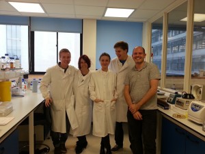
Hi, my name is Claudia Schneider, and my Royal Society University Research Fellowship has allowed me to set up my own group here at ICaMB to study enzymes involved in RNA processing and quality control.
I am originally from Germany, where I did my undergraduate studies and my PhD. Many people might think that Germany is the land of cars or lederhosen – but in truth it is really the land of bread (and beer!). It might therefore not come as a surprise that baker’s yeast has become my favourite model organism.

Click on the image to find out how to make these budding buns!
During my undergraduate studies I was first introduced to RNA and I was (and am still) amazed by its many known and still emerging functions in the cell. We now know that almost the entire eukaryotic genome is transcribed, but only a small fraction of the transcripts are protein-coding messenger RNAs (mRNAs). The others are stable and unstable non-coding RNAs (ncRNAs), which are involved in all aspects of gene expression. RNA molecules are often extensively processed before they’re functional, and each processing step is subject to quality control mechanisms. If you want to know more about the life and death of non-coding RNAs, have a look at this recent review.
Yeast has not always been my first choice to study RNA metabolism, since the object of my PhD project in Prof Reinhard Lührmann’s lab turned out to be completely missing in baker’s yeast. Back then I worked on nuclear pre-mRNA splicing, the removal of non-coding introns from precursor mRNAs catalysed by the spliceosome. It was an exciting time in the splicing field: A second low abundance “minor” spliceosome had just been discovered in most multicellular eukaryotes (with the strange and still not readily explainable exception of C. elegans), and this complex is not present in yeast. The minor spliceosome recognises a rare class of introns (<0.5%) with different consensus sequences at the splice sites and has since been linked to a number of human diseases. During my PhD, I purified and biochemically characterised the snRNP components of this unusual pre-mRNA splicing machinery in human and Drosophila cells.
Since then, I have been fascinated by biochemical and enzymatic assays involving RNA such as in vitro splicing assays, where in vitro transcribed pre-mRNAs are mixed with purified spliceosomes. The goal of such an experiment is to observe precise and (hopefully) pretty intron removal in the test tube – but, to my great annoyance, success was every so often hampered by a powerful ribonuclease (RNase) contamination in the assay that completely trashed the precious RNA substrates. Generic and aggressive RNases like RNase A are found on our skin, are incredibly stable and can even survive boiling.

Common decoration on lab surfaces during my PhD
It is therefore fair to say that my scientific career was majorly influenced by constant warnings by my mentor, who told me that all RNases are evil and must be destroyed. However, for my postdoc, I decided to face my fears and look these evil RNases in the eye, in the humble model system yeast. During my time with Prof David Tollervey at the University of Edinburgh, I learned that there are many different types of RNases, and only very few of them are promiscuous and chop RNA to bits.
The majority of RNases are very sophisticated and versatile enzymes. Several RNases are capable of degrading only specific RNA molecules, or only function under certain circumstances, and protein co-factors often assist in substrate recognition. RNases are crucial elements in RNA quality control or surveillance systems, which distinguish aberrant from “normal” RNA molecules. One clinically important RNA surveillance pathway is called “nonsense-mediated decay” or NMD, and this system recognises and degrades a specific class of defective mRNAs to limit the synthesis of truncated and potentially toxic proteins. NMD defects are linked to ~30% of all inherited human diseases (e.g. Duchenne muscular dystrophy and forms of b-thalassemia) as well as certain types of cancer. In addition to quality control/surveillance, where RNAs are mostly completely degraded, a growing number of RNases have been shown to be responsible for precise processing or “trimming” of precursor RNA molecules to produce their functional forms.
Exonucleases were long believed to be the main players involved in RNA recognition and processing/turnover. However, this model was recently challenged by the identification of endonucleases containing PIN (PilT N-terminus) domains, which appear to play key roles in RNA metabolism. Eight PIN domain proteins and therefore putative endonucleases are encoded in the genome of budding yeast, and this includes three largely uncharacterised “orphan” nucleases.
Overall it is still puzzling to me how individual RNases “make the decision” to either completely degrade or carefully process a specific RNA. Given the ever-growing number of non-coding transcripts in the cell, I am also keen to know which RNases are responsible for which substrates and what the so-far uncharacterised putative PIN domain endonucleases in yeast are doing!
To this end, our lab is using an RNA-protein cross-linking method called “CRAC” (UV cross-linking and analysis of cDNA) to identify the targets of PIN domain endonucleases on a transcriptome-wide scale. The CRAC method and the machinery to cross-link yeast cultures were developed by Sander Granneman at the University of Edinburgh, when we were both PostDocs in Prof David Tollervey’s lab. Sander now has his own lab too, and he runs a CRAC-blog. Interestingly, the cross-linking device we are using was originally designed to sterilise sewage water, but it is now also commercially available for research. With this setup, yeast cells are cross-linked while they are growing in culture, which is crucial to identify the often very transient interactions between nucleases and their target RNAs. This system provides a huge advantage over more traditional cross-linkers like the “Stratalinker”, which requires pelleting and cooling the cells on ice before cross-linking. It is, however, also much bigger and takes up a whole bench in the lab – but I guess there is a drawback to everything! In any case: if you want to find RNA targets for your favourite yeast protein – get in touch!!

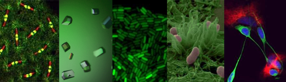
Cultures of Saccharomyces cerevisiae can be “zapped”, while they are growing: It only takes 100 seconds!
Transcriptome-wide RNA-protein interaction analyses generate huge datasets and we use RNA binding and nuclease assays, as well as co-precipitation studies, to validate the in vivo cross-linking results for individual PIN domain endonucleases.
With the help of an ERASMUS exchange student, Franziska Weichmann, who spent 6 months in my lab last year, we have made good progress with two putative PIN domain endonucleases that are linked to ribosome biogenesis. We were able to identify their binding sites on the pre-ribosomal RNAs, as well as co-factors that are important to recruit them into the pre-ribosome. We have also set up an in vitro system with recombinant proteins, and we are currently trying to convince one of them to specifically cleave its proposed rRNA substrate in the test tube – and we are slowly getting there…..
Like the other ICaMB ECRs, who posted on this Blog before, I would like to finish by saying that having my own lab has been an exciting and (on most days) enjoyable adventure so far – and I am looking forward to the next set of challenges…
 In the latest of our series focussing on the Early Career Researchers (ECRs) in ICaMB, we feature