ECRs at ICaMB: For he’s a jolly good (RS) Fellow

As you saw from our post a couple of weeks ago, ICaMB has recruited several new PIs. To make sure we all know more about them and their research, we decided to have a series specially dedicated to them: Early Career Researchers (ERCs) at ICaMB. First in the series is Dr Kevin Waldron.

by Dr Kevin Waldron

As some of you will already know, I was awarded a Sir Henry Dale fellowship in 2012. This is a new funding scheme for early career researchers, which is co-funded by the Wellcome Trust and the Royal Society. The scheme provides exceptional research support for fellows, including funding a postdoctoral research assistant for the duration of the project (5 years initially, with potential extension for a further 3) as well as all direct research costs, with the aim of supporting the researcher while they forge an independent research career and carve out their research niche. As a result they are highly coveted and extremely competitive – the interview at the Wellcome Trust in July 2012 was probably the most intimidating experience of my career. My award was one of the very first round, in which 10 awards were given from a pool of about 100 applications.

Enjoying the sights of Ha Long Bay (Vietnam)… and doing some deep thinking about my science, of course…

Though the grant was awarded in Summer 2012, I only activated the grant in May 2013 (primarily so that I could swan off on an extended holiday travelling round South East Asia – but that’s another story). Since May there have been two new additions to my lab team; Emma Tarrant, a postdoctoral research associate funded by the fellowship grant, and Anna Barwinska-Sendra, a new PhD student. This combination of good funding and great new people will allow us to really get our teeth into our research question over the coming years, free of the hassle of grant writing and worrying about where the next funds are coming from.


I have been studying bacterial metal homeostasis since I started my PhD with Prof. Nigel Robinson (now in Durham), way back in 2003. During that period I studied cyanobacteria, but my current research is focused on the Gram positive mammalian pathogen Staphylococcus aureus. S. aureus has become a major medical problem in recent decades due to the rise of strains that are resistant to multiple conventional antibiotics, with the term “superbug” and the acronym “MRSA” entering the popular consciousness – see for example here, here and here. The organism is particularly problematic in hospitals, where it causes significant morbidity and mortality (see for example these UK and US studies). Though infection control measures introduced in hospitals are proving successful in reducing the number of S. aureus-related deaths, both the prevention and treatment of hospital-acquired infections remain major burdens on patients and on the NHS budget. S. aureus is also problematic in farm animals, being a major cause of bovine mastitis and of lameness among broiler chickens.
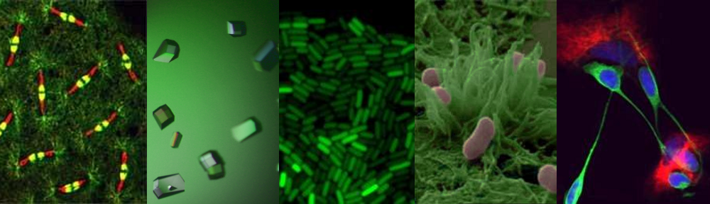
My favourite metal

One intriguing new approach to prevention of infection in such settings is the use of solid copper or copper-containing alloys. Copper surfaces show wide-spectrum antimicrobial activity and are certified by the US EPA as antibacterial and ‘self-sanitising’. Multiple trials have shown that these materials can reduce bacterial copper load on touch surfaces in hospitals (see here for example), and the effects on disease transmission are currently being tested. In fact, this is not a new approach at all: as this YouTube clip explains, copper has been used since ancient times to sterilise drinking water and to treat minor ailments, even being mentioned by Hippocrates in ~400 BC. Copper is the active ingredient in numerous agricultural fungicides including Bordeaux mixture, in use since 1885.

The mechanisms by which copper, either as metal salts or as solid metal surfaces, kills bacteria are unknown. Dissolved copper ions are thought to play an important role in the killing mechanism even from solid copper surfaces. In fact, the toxicity of excess concentrations of essential metal ions (as opposed to non-essential metals such as lead and mercury) has been historically under-studied and has merely been seen as a confounding factor of studies of normal metal homeostasis. Copper is redox-active, a potent Fenton catalyst, and therefore may catalyse the production of reactive oxygen species in vivo. It is also at the top of the Irving-Williams series, meaning it will bind extremely tightly to proteins, potentially having deleterious effects on their function when present in excess.

Our goal is to identify and understand these toxicity mechanisms, both to shed light on new aspects of metal homeostasis and to find better ways of exploiting this toxicity for medical and commercial applications. To do this we’re going to use a combination of multiple experimental approaches in collaboration with a number of world experts in diverse fields. We will use proteomic methods to identify S. aureus proteins that become aberrantly associated with copper under high-copper growth conditions, and then characterise the functional effects of those aberrant associations using traditional biochemistry and molecular genetics. We will combine this with metabolomics studies of the effects of copper toxicity on bacterial metabolism. Finally, we will use genetic screens to identify mutant strains that display increased copper resistance. Together, this should give us a comprehensive view of the effects of copper toxicity on the cell, with the aim of elucidating the multiple mechanisms by which excess copper interferes with normal function.

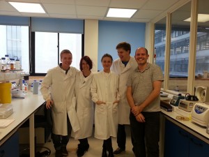
The Waldron lab group: Jack Stevenson, Emma Tarrant, Anna Barwinska-Sendra, Stuart York and Kevin Waldron (left to right)


So we’re just starting off on a long-term quest, with lots of experiments to keep us busy for the next few years. It’s an exciting time in the Waldron lab.


Links

Sir Henry Dale Fellowships   http://www.wellcome.ac.uk/Funding/Biomedical-science/Funding-schemes/Fellowships/Basic-biomedical-fellowships/WTDV031823.htm
Wellcome Trust www.wellcome.ac.uk/index.htm
Royal Society www.royalsociety.org
US EPA www.epa.gov/pesticides/factsheets/copper-alloy-products.htm

A ‘tail’ to tell – Lakey lab discovery could lead to a new class of antibiotics


Dr Chris Johnson

“Most of us know that we should wash our hands after being around animals but do most of us know the reasons why? As a researcher who spends most of his time in the lab killing E. coli, using E. coli specific antibiotics I should be well aware of the dangers of this often underestimated Gram-negative bacteria. However when my youngest daughter contracted E. coli O157 after visiting an agricultural show in Scotland in 2011 (even though we had followed the hand washing procedure), I realised that I did not appreciate how nasty this bacteria can be. Thankfully after 6 weeks in hospital including a lengthy stretch on dialysis my daughter made a full recovery but not everyone is so lucky.”

Professor Jeremy Lakey

The urgent need to develop new drugs to target pathogenic bacteria has been a theme of the ICaMB blog since its inception in early 2013.  However, these words from Dr Chris Johnson, a postdoc in Jeremy Lakey’s laboratory, bring home the seriousness of the problem.  Fortunately, Chris is in a position to do something about this.

Many of you will have realised that last week saw another potential breakthrough in ICaMB’s ongoing ‘War on bacteria’ (and here, here and here).   This time the PI making the news, with data demonstrating the possibility of a whole new class of antibiotics, was Professor Jeremy Lakey.  And when we say making the news, we mean that literally. The ITV interview of Jeremy and his team can be viewed here

This story has made news around the world and has been featured in newspapers in Australia and India (and here), as well as closer to home.  Here is the official university press release.

So what’s going on?  If you want to read the primary paper it is here

The unstructured domain of colicin N kills Escherichia coli. Mol Micro 89:84-95

Chris Johnson, who was the lead author on this manuscript explains:

E. coli produces protein antibiotics called colicins which are used to kill E. coli and closely  related bacteria in the eternal bacterial arms race. In order to further understand how one of these, colicin N (ColN), works, we dissected the protein into its individual domains to see how each part behaved in isolation. Quite by chance we found that part of the protein, THE TAIL, was actually toxic to them. Although far less efficient than the entire ColN molecule, it remains specific for E. coli and furthermore, the specificity is housed within an intrinsically unfolded domain (a domain which has no defined 3D structure). Although this is a very basic discovery in its early stages, it allows us to appreciate novel mechanisms to kill E. coli.” (see the full version of this at the bottom of the page)

Some more about Jeremy

Any of you who were bioscience undergraduates in Newcastle will know Jeremy Lakey from his famous recreations of protein structure using party balloons.  Others may know him for co-founding Orla Protein technologies.  Some of us know him as a man who will always buy his round in the pub.  All of us know him as a great scientist and colleague.  Furthermore, Jeremy is a leading supporter of Leading Edge and recently a group of 6 Year 9 school pupils from St Cuthberts RC School looked at how ColN acts against E.coli when you start changing the amount of salt they are grown in. As many ICaMB scientists may know Jeremy also runs a workshop with Ponteland Community High School to explore bacterial shape and their surfaces using LEGO.

But did any of us think that one day Jeremy Lakey may SAVE THE WORLD from antibiotic resistant bacteria? Possibly.

The detailed science

Most Gram- negative bacteria produce protein antibiotics which are used as weapons in the battle between competing populations of bacteria.  E. coli produces protein antibiotics called colicins which are use to kill E. coli and closely related bacteria.  Once colicins are released into the extracellular milleu they dock onto their targets via specific outer-membrane receptors and then seek out an internal, periplasmic, binding partner (the Tol or Ton proteins) which helps them translocate into the cell.  We study colicin N (ColN) which comprises of an intrinsically unfolded N-terminal translocation (T) domain, involved in TolA and OmpF binding.  Its central receptor binding (R) domain binds lipopolysaccharide whilst its C-terminal 200 amino acids define the cytotoxic pore-forming (P) domain. This latter feature is common to all pore-forming colicins and forms a channel in the inner-membrane causing K+ release and cell death.  Other colicins have C-terminal domains which display cytotoxic activities that include DNAase or RNase activity.  Irrespective of the particular cytotoxic activity, all colicins are comprised of three domains (T-R-P) and it was assumed that the sole role of the T and R domains was to deliver the cytotoxic C-terminal domain across the outer-membrane.

In order to investigate the mechanism of ColN activity we dissected the protein into its individual domains.  We were attempting to block the toxic activity of full length ColN by pre- incubating E. coli cells with the intrinsically unfolded T-domain.  The rationale behind the experiment was that we could block all the available receptor sites on the E. coli target cells by saturating with T-domain, such that when the full length ColN was added to cells it would be non-toxic, as all the essential receptor binding sites would be already sequestered.  However rather than protecting the cells, T-domain was found to be toxic and like full length ColN provoked K+ efflux.   Although less efficient than full length ColN, T-domain is strictly dependent upon the same receptor proteins, OmpF and TolA for killing.  Since these receptors are only found in E. coli-like bacteria, T-domain displays the unusual combination of a generic killing mechanism coupled with extreme specificity housed within an intrinsically unfolded domain.

Links

Jeremy Lakey’s University home pagehttp://www.ncl.ac.uk/camb/staff/profile/jeremy.lakey

Follow Jeremy Lakey on Twitterhttps://twitter.com/JeremyLakey

ICaMBhttp://www.ncl.ac.uk/camb/

The Centre for Bacterial cell Biologyhttp://www.ncl.ac.uk/cbcb/

The official Newcastle University press release: http://www.ncl.ac.uk/press.office/press.release/item/chance-finding-could-lead-to-new-antibiotics

Link to ITV story:

http://www.itv.com/news/tyne-tees/story/2013-07-05/breakthrough-in-combatting-bacterial-infection/

The ‘Australian’ storyhttp://www.theaustralian.com.au/news/breaking-news/tail-could-be-used-for-new-drugs/story-fn3dxix6-1226674131684

The ‘Times of India’ Storyhttp://articles.timesofindia.indiatimes.com/2013-07-06/science/40406695_1_escherichia-coli-e-coli-protein

The Northern Echo storyhttp://www.thenorthernecho.co.uk/news/10524003.Scientists__chance_find_may_develop_new_generation_of_antibiotics/

Jeremy’s company, Orla Proteinshttp://www.orlaproteins.com/about-orla/the-board.aspx


Breaking news: Mitochondria replacement therapy given green light

Breaking news by Prof Bob Lightowlers

Professor Dame Sally Davies, the Governments Chief Medical Officer, has today given support to a change in the law that will allow the form of mitochondrial replacement therapy that is being pioneered here in Newcastle, has we mentioned before.

Its great news for any prospective mother with mitochondrial DNA disease who is concerned about transmitting the disease to their children.

Most media is covering this – BBC, Guardian, Telegraph – but if you are still confused, we thought it would be good to recap the essential points:

Electron micrograph of a cell (coloured blue) revealing part of the mitochondrial structure (orange) within. The entire length of the mitochondrion is about 5 micrometres.

– Mitochondria are crucial structures found in all cells in our body and they take our common foodstuffs such as fats and sugars and turn them into energy. They have their own genetic element, mitochondrial (mt) DNA. Much smaller than our chromosomes, mtDNA is essential for energy production.

– In 1988, scientists in the UK and US recognised that certain diseases were caused by mutations in mtDNA with the main disorders related to your muscle tissue and the brain.

– It is estimated that at least 1:10,000 people suffer from disorders associated with defects in mtDNA – that’s more than 6,000 people in the UK.

– Mitochondrial DNA is only transmitted to babies by their mothers. Unfortunately, as you inherit your mothers mitochondria, diseases caused by mtDNA mutations are inadvertently transmitted from the mother.

– Doug Turnbull, Director of the Wellcome Trust Centre for Mitochondrial Research (WTCMR) and I have wondered since the 1990s whether it would be possible to prevent the transmission of the faulty mtDNA from the mothers to their children by transferring the nucleous from an egg of the mother carrying these mtDNA mutations into a healthy egg whose nucleous had been removed, and then fertilise it

– Professor Mary Herbert, Doug and a group of us from the Mitochondrial Research Group were able to show that such a swap could be performed without any or very low levels of the defective mtDNA being transferred. Importantly, there was also no defect detectable in the reconstituted cells.

In August 2012, the government asked the Human Fertility and Embryological Authority (HFEA) to find out what the general public thought of the procedure.  The results were collated  and the Human Fertility and Embryological Authority made a recommendation to Government. There was an overall support for the new technology with only 10% being fairly or strongly against the concept of mitochondrial gene replacement

To understand all the details and find out the whole story, see my post a while ago here.

The Wellcome Trust also has a very nice video explaining the science behind the news story:


Links

Wellcome Trust Centre for Mitochondrial Research http://www.newcastle-mitochondria.com/
Human Fertility and Embryological Authority (HFEA) http://www.hfea.gov.uk/index.html
HFEA mitochondria puclib consultation 2012 http://www.hfea.gov.uk/6896.html


ICaMB Research Update: Zenkin lab Science paper

Congratulations to ICaMB and CBCB researchers Soren Nielsen, Yulia Yezenkova and Nikolay Zenkin and  who have a paper published in the prestigious journal Science today.

Nikolay Zenkin: RNA Polymerase researcher

There are three RNA Polymerases in Eukaryotic cells and although much attention focuses on the role of RNA Polymerase II, since it transcribes mRNAs from protein encoding genes, it is easy to forget that the majority of transcription in the cell is accomplished by RNA Polymerase III.  RNA Polymerase III (or Pol III as it is commonly known) is responsible for the synthesis of ribosomal 5S rRNA, tRNA and other small RNAs.


Soren’s paper, entitled “Mechanism of Eukaryotic RNA polymerase III Transcription” termination solves a long-standing mystery in the field of how transcriptional termination by RNA polymerase III takes place. Their study reveals an elegant scenario, in which co-transcriptional folding of highly-structured RNA polymerase III transcripts causes termination at the end of their genes. This mechanism ensures proper folding of the structural/catalytic RNAs synthesized by RNA polymerase III prior to RNA release. Analogies with bacterial termination suggest that this fundamental mechanism may have emerged before divergence of bacteria and eukaryotes.

Here is the Science Editor’s summary of the paper

It is as important to terminate any biological process as it is to start it. Transcription, copying information encoded in genes into RNA, requires accurate and timely termination. Nielsen et al. (p. 1577) present a mechanism for transcription termination by RNA polymerase III, the enzyme that synthesizes the majority of RNA molecules in eukaryotes. In this scenario, the folding of the RNA as it is transcribed by polymerase into a highly structured transcript causes termination at the end of its synthesis. This mechanism may serve as a control of proper folding of structural or catalytic RNAs synthesized by RNA polymerase III. Comparison with other organisms suggests that this mechanism emerged before divergence of bacteria and eukaryotes.

And here is the abstract of their paper

Gene expression in organisms involves many factors and is tightly controlled. Although much is known about the initial phase of transcription by RNA polymerase III (Pol III), the enzyme that synthesizes the majority of RNA molecules in eukaryotic cells, termination is poorly understood. Here, we show that the extensive structure of Pol III–synthesized transcripts dictates the release of elongation complexes at the end of genes. The poly-T termination signal, which does not cause termination in itself, causes catalytic inactivation and backtracking of Pol III, thus committing the enzyme to termination and transporting it to the nearest RNA secondary structure, which facilitates Pol III release. Similarity between termination mechanisms of Pol III and bacterial RNA polymerase suggests that hairpin-dependent termination may date back to the common ancestor of multisubunit RNA polymerases.

Links

Link to the Science paper: http://www.sciencemag.org/content/340/6140/1577.abstract

Nikolay Zenkin laboratory home page: http://www.ncl.ac.uk/camb/staff/profile/nikolay.zenkin#tab_research

Centre for Bacterial Cell Biology: http://www.ncl.ac.uk/cbcb/

Science magazine: http://www.sciencemag.org/

Link to the Science cover: http://www.sciencemag.org/content/340/6140.cover-expansion

You don’t always want what your mother gives you! – can we prevent mitochondrial disease?


By Professor Robert Lightowlers

In 1988, scientists in the UK and US recognised that certain diseases were caused by mutations in mtDNA . Over the following 20 years, mtDNA defects have been shown to cause a range of debilitating diseases many affecting different parts of the body. However, the main disorders relate to your muscle tissue and the brain.

Human muscle fibres stained for mitochondrial function. As can be seen in B, some of the fibres show no activity. This is because these fibres have high levels of mutated mitochondrial DNA.

It is estimated that at least 1:10,000 people suffer from disorders associated with defects in Mitochondrial DNA (mtDNA) – that’s more than 6,000 people in the UK. Even so, it is only recently that the importance of mitochondrial diseases have hit the general media.

Many of you will have seen the debate on correcting mitochondrial diseases in the newspapers (for example, see the Guardian, Telegraph) and on television recently, but not be aware of the central role that Newcastle researchers have played in making this exciting, or to some, controversial, new therapy closer to becoming a reality.  Here, Bob Lightowlers ICAMB Director and senior member of the Wellcome Trust Centre for Mitochondrial Research (WTCMR) reflects on the role mitochondrial research in Newcastle has played in this process over the last 20 years and tells us some of the story behind the headlines.

What are Mitochondria?

Electron micrograph of a cell (coloured blue) revealing part of the mitochondrial structure (orange) within. The entire length of the mitochondrion is about 5 micrometres.


These crucial structures found in all the trillions of cells in our body have many essential functions. One very important role they play is to take our common foodstuffs such as fats and sugars and turn them into energy for our body’s to function.

A single human cell showing the nucleus (green), the mitochondrial network (red) and the mitochondrial DNA within the network (yellow)


One surprising element of these structures is that they contain their own genetic element, mitochondrial (mt) DNA. Much smaller than our chromosomes, mtDNA is essential for energy production.


OK, so this is important, but why have mitochondria and mtDNA begun to work their way into the common conversation of the nation?

Answer: Our mothers!

What has this got to do with our mothers ? Mitochondrial DNA is only transmitted to babies by their mothers. This is different to all our other DNA where copies are made and transmitted from both parents. Unfortunately, as you inherit your mothers mitochondria, diseases caused by mtDNA mutations are inadvertently transmitted from the mother.

How does this relate to Newcastle based Mitochondrial Research?

My colleague Doug Turnbull, a neurologist here in Newcastle (and Director of the WTCMR) and I have been intrigued by these mtDNA mutations since it first became clear that they could cause disease. Back in the early ‘90’s, we discussed whether some day it would be possible to try and prevent the transmission of the faulty mtDNA from the mothers to their children. Of course, at that stage, it was just wishful thinking. As the Mitochondrial Research Group (MRG) began to grow and mature in Newcastle, we often returned to one question:

What if the nucleus from the diseased egg could be transferred to a healthy egg whose nucleus had been removed, in essence leaving all the affected mtDNA behind ?

If it was indeed possible, this reconstituted egg could be fertilised and implanted back into the mother by standard techniques used routinely in fertility clinics throughout the world. We also would consider when would such a technique be most efficient: before or after fertilisation of the egg? On paper both options looked possible, but there are many complications.

Technical Concept: achieving the switch of nuclei without some of the faulty mtDNA being inadvertently taken along for the ride.

Towards the end of the 90’s, scientists working in Canada were able to show that the level of mtDNA inadvertently transferred when the nucleus was switched into a recipient cell lacking a nucleus, was low. This was a promising result, but it led to two central questions:

•   Could this be repeated with human cells?

•   Was this technique morally and ethically acceptable to everyone?

The ethical debate: Debate raged as to whether this technique would constitute genetic manipulation of humans, which of course would be illegal. Further, it was not possible to perform these types of reconstruction experiments in man, as using viable human fertilised cells for research was also, understandably, illegal.

Professor Mary Herbert working at the nearby Human Fertility Centre came up with an intriguing proposal. She explained that unfortunately, during the standard process of in vitro fertilisation, many eggs became incorrectly fertilised. These eggs are unable to grow correctly and have to be discarded. One way of determining whether it would be possible to swap mtDNA in humans, she suggested, was to use these incorrectly fertilised eggs. As this procedure would still require the manipulation of fertilised human eggs, a licence would need to be applied for from the Human Fertilisation and Embryological Authority (HFEA). Following lengthy and extensive debate, including members of the research team being called to the House of Commons, a licence was eventually awarded in 2005.  Five years later, with the essential help of colleagues in the Fertility Centre, Mary, Doug and a group of us from the MRG were able to show that such a swap could be performed without any or very low levels of the defective mtDNA being transferred. Importantly, there was also no defect detectable in the reconstituted cells . In 2011, this very promising result, along with many other important contributions made by the Newcastle MRG to understanding mitochondrial biology in health and disease was recognised by the Wellcome Trust who funded the establishment of a new Research Centre in Newcastle, the Wellcome Trust Centre for Mitochondrial Research.

Getting acceptance of the technique

It was important to know whether the people of the UK agreed that such reconstitution technology was ethically acceptable. In August 2012, the government asked the Human Fertility and Embryological Authority (HFEA) to find out what the general public thought of the procedure .  The results were collated last month and the Human Fertility and Embryological Authority made a recommendation to Government. There was an overall support for the new technology with only 10% being fairly or strongly against the concept of mitochondrial gene replacement ) This is an endorsement of the method but there is still a long way to go before the technique can be performed in the clinic.

Its amazing to think how far this concept has come in 20 years. Perhaps in another 20 years we may be able to look back and celebrate how this dream has helped to provide a realistic method to help prevent the transmission of a debilitating disease for many couples.


Wellcome Trust Centre for Mitochondrial Research http://www.newcastle-mitochondria.com/
Human Fertility and Embryological Authority (HFEA) http://www.hfea.gov.uk/index.html
HFEA mitochondria puclib consultation 2012 http://www.hfea.gov.uk/6896.html

Bulging bacteria and the origins of life

Jeff (left), Romain (centre) and Yoshikazu (right), the team of researchers behind these exciting discoveries


In a paper published this week in CellJeff Errington’s team in ICaMB, have discovered new insights into the origin of life on Earth.


Jeff and his team share their results

Bacteria were the first organisms to appear on planet earth. Almost all modern bacteria have a tough protective shell called a cell wall. The structure of the wall and the mechanisms used by cells to manufacture it are conserved, suggesting that the wall was invented right at the beginning of bacterial evolution, and, therefore, when the first true cells emerged.

Production of cell wall is carefully regulated by complex machineries that allow the cell to enlarge and then divide in a controlled manner, all the time maintaining the integrity of the wall intact.

Despite its importance, it seems that many modern bacteria can survive cell wall loss under certain very special conditions, such as when they are treated with certain antibiotics that interfere with its production, like penicillin. Not only that, but a few years ago my lab showed that these “L-form” cells (named after the Lister Institute in London where they were first described) no longer need the complex mechanisms normally needed for bacterial growth and division. Instead, they grow by extrusion of irregular tubes or blebs of cytoplasm, that pinch off into daughter cells.

Our team – me, Yoshikazu KawaiRomain Mercier – has been working on this problem for some time. “Studying L-form biology is a real technical challenge, and this work could not have succeeded without the strong collaboration established between us“, says Romain. As Yoshikazu explains: “we developed a very simple genetic system to isolate mutations enabling L-form development from non-viable protoplasts.

We are excited because we think we have now solved the mystery of how L-forms grow and divide. Our latest results, published in Cell, show that the mechanism is remarkably simple: it requires only that cells make excess amounts of membrane – the thin porous layer that acts as the outer boundary of all cells, including our own.

Increasing the membrane surface area beyond the amount needed to contain the cytoplasm causes the cell to buckle and distort. Eventually, this leads to pinching off of membrane bags that are ill formed but nonetheless viable “baby” cells.

Time-lapse photography representing the division of B. subtilis without cell wall (L-form). The images were obtained using light microscopy. Scale bar: 3 μm

At first, we thought this mechanism was too simple to be true, we changed our minds when we were alerted to amazing experiments being done by several groups working on the origins of life, particularly Jack Szostak at Harvard, Saša Svetina in Ljubljana and Peter Walde in Zurich. These groups have been wondering how primitive cells could have arranged to grow and divide efficiently without spilling all of their contents. They recently found that simple membrane bags, called “vesicles”, can be induced to grow and reproduce into multiple smaller vesicles, in the test tube, just by increasing their surface area.

So, in explaining how the bizarre L-form bacteria manage to survive the loss of their beloved cell wall, we think we may now also have glimpsed how the first primitive cells could have duplicated themselves at the dawn of life on earth.

Jeff Errington 
Director of the Centre for Bacterial Cell Biology


Cell paper: http://www.cell.com/abstract/S0092-8674(13)00135-9
Cell website: http://www.cell.com/home see PaperFlick
Newcastle University Press Release:http://www.ncl.ac.uk/press.office/press.release/item/how-did-early-primordial-cells-evolve#.US-chen77jQ

Soapbox Science guest blogpost: http://www.blogs.nature.com/soapboxscience/2013/02/28/social-media-from-an-institutional-perspective-why-are-we-on-there

ICaMB website: http://www.ncl.ac.uk/camb/
Facebook: https://www.facebook.com/pages/ICaMB-Newcastle/416200498466481
Twitter: https://twitter.com/ICaMB_NCL
YouTube: http://www.youtube.com/channel/UCSuZgA6URiXTUoHq1tMe-PQ