How a motile cytoskeleton drives bacterial cell division

seamusIn a recent issue of Science, the discovery of a key mechanism for bacterial cell division was reported. This work was carried out by Dr Seamus Holden’s lab (ICaMB) in collaboration with Professor Cees Dekker (TU Delft), Professor Yves Brun (Indiana University), Professor Mike VanNieuwenhze (Indiana University) and Professor Ethan Garner (Harvard University). Here, Seamus tells us about this discovery and what its implications could be for antimicrobial research.

Bacterial cell division is a lovely mechanistic problem in biology: how do the simplest living organisms build a crosswall at mid cell, against very high outwards pressure (think of a racing bike tyre), without bursting?  A ring of protein filaments forms around the future division site, and enzymes associated with this ring build a new crosswall that cleaves the bacteria in half. But what has remained completely mysterious is how these proteins work together as a single nanoscale machine to cut the bacterial balloon skin (cell wall) in two.

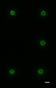
Cytoskeletal proteins FtsZ in live bacteria imaged in vertical nanocages

Cytoskeletal proteins FtsZ in live bacteria imaged in vertical nanocages

Working together with collaborators in Delft, Indiana and Harvard, we tracked the organization and motion of key division proteins as they build the dividing crosswall, and the organization of the newly built crosswall itself. We began by examining the motion of FtsZ, a cytoskeletal filament that is required for cell division – cytokinesis – in bacteria and is related to the tubulin cytoskeletal protein found in eukaryotic cells. Using high-resolution microscopy techniques, we found that FtsZ filaments move around the division site, traveling around the division ring. We imaged the motion of individual cell wall synthesis enzymes, and saw that the synthesis enzymes ride on FtsZ filaments, building new cell wall as they travel along the division site. This causes the cell wall to be synthesized in discrete sites that travel around the division site during cytokinesis, a process which we were able to observe directly by using dyes that label the bacterial cell wall. Using a variety of experimental techniques, we were able to speed up or slow down how fast FtsZ rotated around the cell. Strikingly, we found that the speed of FtsZ filament motion determines how fast the cell can divide. When FtsZ moves more rapidly, cell wall is produced more quickly, and cytokinesis happens faster. This shows that the motion of FtsZ is the critical overall controller of cell division.

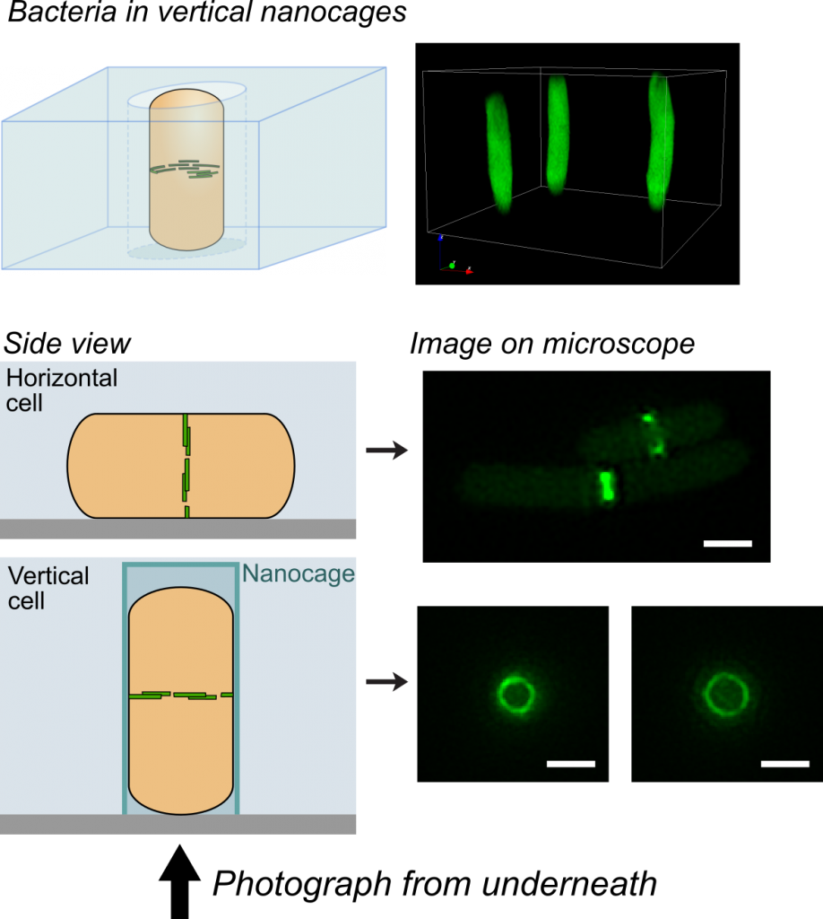
One challenge that we faced was trying to look at the division proteins in actively dividing cells. At the earliest stages of division, it was possible to image division protein organization because the proteins in the partially assembled ring are sparsely distributed. However, a new strategy was required to measure how the dense protein network of actively dividing cells was organized. Normally, bacteria are immobilized flat on a microscope slide, and imaged from underneath, but unfortunately this places the division ring side-on, obscuring the motion and organization of division proteins. To solve this problem, we used nanofabrication technology, originally developed to manufacture computer chips, to create tiny gel nanocages to trap bacteria in an upright position.

Bacteria trapped in vertical nanocages

Bacteria trapped in vertical nanocages

By trapping individual bacteria upright, we were able to rotate the cell division ring so that it was fully visible on our high resolution microscope. This revealed the dynamic motion of FtsZ filaments as they travel around the entire division site:

Together, these results revealed the basic mechanistic principles of bacterial cell division: that the building of the division crosswall is orchestrated by moving cytoskeletal filaments.  Previously, the cytoskeleton was thought to serve as a static scaffold, recruiting other molecules and perhaps exerting some force to divide the cell. This new work demonstrates that all the components of cell division are in constant, controlled motion around the division site, driven by the fundamental dynamics of the cytoskeleton.

In the longer term, this study could open up novel antibiotic targets. Based on the discovery that the treadmilling motion of the bacterial cytoskeleton is critical for division, it may be possible to develop new drugs that specifically inhibit this motion, similar to how the chemotherapy drug taxol suppresses the motion of the cytoskeleton in cancer cells.

==================================

Links

Explanatory animation: https://youtu.be/6dq2_gqKPfU (Animation credit TU Delft / Scixel)

Nanocage Video: nanocage-movie-2.

Science report: http://science.sciencemag.org/content/355/6326/739

Press release: http://bit.ly/2kwsnJf

A novel copper storage protein discovered by ICaMB scientists

In a recent issue of Nature the discovery of a new family of proteins that store copper ions inside bacterial cells was reported. This work was carried out by Prof. Chris Dennison’s lab (ICaMB) in collaboration with Dr. Kevin Waldron (ICaMB), Dr. Arnaud Baslé (ICaMB), Dr. Neil Paterson (Diamond Light Source) and Prof. Colin Murrell (UEA). Here, Kevin tells us about this discovery and what its implications could be for the biotechnological exploitation of methane.

Methane, the main component of natural gas, is produced by both geological and anthropogenic processes on Earth, and can be released into the atmosphere. The biosphere’s primary mechanism for limiting the amount of methane (a powerful ‘greenhouse gas’) that escapes into the atmosphere is through its consumption by methane-oxidising bacteria, or methanotrophs (literally ‘methane-eating’), who utilise methane as both a carbon and energy source.

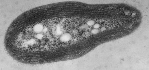
Transmission electron micrograph of a M. trichosporium OB3b cell showing the intracytoplasmic membranes that house the copper-dependent enzyme pMMO.

Transmission electron micrograph of a M. trichosporium OB3b cell showing the intracytoplasmic membranes that house the copper-dependent enzyme pMMO.

The key enzyme that methanotrophs use to oxidise methane to produce methanol is called methane monooxygenase (MMO). Almost all methanotrophs possess a membrane-bound form of MMO that requires copper (Cu) for activity, called pMMO (some strains also possess an iron-requiring alternative soluble enzyme, called sMMO). Therefore, these bacteria have an unusually high demand for copper, a metal that is essential for most organisms but is used rather sparingly, mainly because it can also be extremely toxic.

Previous studies of methanotrophs have revealed that they utilise unusual mechanisms for acquiring the copper they need for pMMO. For example, they produce and secrete a small, modified peptide called methanobactin that is involved in high affinity copper uptake, analogous to the more familiar siderophore iron uptake systems.

The model methanotroph, Methylosinus trichosporium OB3b, was analysed in the hope of uncovering novel copper proteins. Soluble extracts were resolved by two-dimensional liquid chromatography, with the copper content of resulting fractions monitored. The most abundant copper pool was found to contain a small, cysteine-rich protein that had not been studied previously and Chris Dennison (PI) and I (Co-I) obtained a BBSRC grant to investigate this and related proteins.

The presence of 13 cysteine residues in a mature protein (after cleavage of its signal peptide) with only 122 amino acids was immediately significant, as its thiol sidechain is often found in copper-binding sites. The recombinant protein was extensively studied using a suite of biochemical and biophysical methods to determine its in vitro properties This novel tetrameric protein is capable of binding up to 52 Cu(I) ions with high affinity, consistent with a potential role in the storage of this essential metal and was therefore named Csp1 (copper storage protein 1). A mutant strain of M. trichosporium OB3b that lacks this Csp1 protein (as well as a closely-related protein, Csp2) shows a phenotype consistent with a copper storage role of these proteins in vivo.

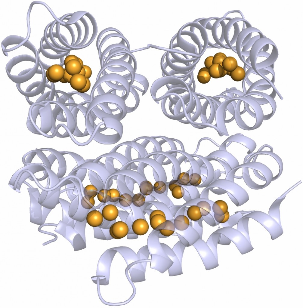
The crystal structure shows that the apo-monomer adopts a well-characterised four-helix bundle motif with all 13 cysteine sidechains pointing into the core of the bundle. The protein structure is essentially unchanged in the presence of copper, except that the core of each monomer has filled with 13 Cu(I) ions, bound by the cysteine sidechains. This is an unprecedented structure for metal storage.

The structure of the Cu(I)-Csp1 tetramer, with the 13 Cu(I) ions bound within the core of each four-helix bundle shown as orange spheres.

The structure of the Cu(I)-Csp1 tetramer, with the 13 Cu(I) ions bound within the core of each four-helix bundle shown as orange spheres.

The implications of this discovery could be of wide impact. There is a great deal of interest in using methanotrophs and MMOs in industrial biotechnology. Available methane is on the increase due to ‘fracking’, but importantly methane can also be produced from sustainable sources such as through the degradation of biomatter (biogas). Methanotrophs and the MMOs could in future be exploited using synthetic biology approaches for gas-to-liquid conversion, for the production of liquid fuels as well as bulk and fine chemicals from renewable methane.

Any such biotechnological applications require an understanding of how the production of pMMO is regulated and how this complex enzyme is assembled, including the cellular delivery and insertion of the essential copper cofactor. The work published in Nature shows that Csps are an important piece in that assembly jigsaw.

Furthermore, bioinformatics has shown that cytosolic members of the Csp family are also encoded in other bacterial genomes. Most bacteria are thought not to require copper in their cytoplasm, so the presence of cytosolic Csp homologues in their genomes could re-write our basic understanding of their use and homeostasis of copper.

Links:

Nature article: http://www.nature.com/nature/journal/v525/n7567/full/nature14854.html

News & Views: http://www.nature.com/nchembio/journal/v11/n10/full/nchembio.1918.html

BBSRC website: http://www.bbsrc.ac.uk/news/fundamental-bioscience/2015/150827-pr-bacteria-that-could-protect-our-environment/

ACS Chemical & Engineering news: http://cen.acs.org/articles/93/web/2015/08/Protein-Stores-Copper-Methane-Digesting.html

Press release: http://www.ncl.ac.uk/press.office/press.release/item/could-bacteria-help-protect-our-environment

Beer, Bread and Bacteria


Prof. Harry Gilbert

Prof. Harry Gilbert

The diverse organisms that live in our gut, collectively termed the gut microbiota, can have a major impact on our health. A new paper from Prof. Harry Gilbert’s lab in ICaMB, published today in Nature, shows that one member of our gut microflora uses a ’selfish’ mechanism to degrade a component of the cell wall of the yeast in our diet.

By Drs. Elisabeth Lowe and Fiona Cuskin.

Have you over-indulged this Christmas? One too many ales, or too many turkey sandwiches?

Beer: full of yeast.

Beer: full of yeast.

The good news is that, while your waist line may be expanding and you may be feeling a little the worse for wear, the organisms of your gut microbiota are thriving!

Some of the bacteria in the human gut, called Bacteroides, can break down the complex carbohydrates in our diet into simple sugars, which they use for energy.

Bread: full of yeast


Our work, published in Nature today, shows that one of these bacteria can also break down some of the carbohydrate cell wall of yeasts, called mannan, a polymer of mannose. This includes mannan from Saccharomyces cerevisiae (Baker’s yeast), and the pathogenic gut fungus, Candida albicans. The current understanding of polysaccharide digestion by gut bacteria suggests a cooperative environment, in which break-down products are shared between different members of the microbiota. However our data shows that this species (Bacteroides thetaiotaomicron) is ‘selfish’ when it degrades mannan, and doesn’t share any of the digestion products with other bacteria.

Turkey: er... full of yeast?

Turkey: er… full of yeast?

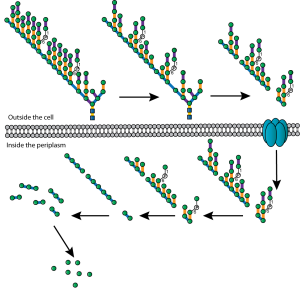
To achieve this selfish degradation, the large and multiply-branched mannan structure undergoes only very limited degradation in the extracellular space (see figure). The resulting processed mannan fragments are then imported into the Bacteroides periplasmic space, where they are further degraded by a suite of glycoside hydrolase enzymes to yield the sugar mannose. Achieving complete breakdown within the periplasm prevents  the mannose from being shared with competing bacteria.

This selfish approach to mannan utilisation could offer the opportunity to design bespoke prebiotics targeted to a specific bacterial population within the gut. Food products containing yeast mannan would be expected to promote growth of B. thetaiotamicron, whereas other complex polysaccharides may specifically promote other members of the microbiota.

Complex polysaccharide mannan is sequentially degraded by a 'selfish' mechanism to yield periplasmic mannose.

Complex polysaccharide mannan is sequentially degraded by a ‘selfish’ mechanism to yield periplasmic mannose.

A number of inflammatory bowel diseases such as Crohn’s disease are associated with intolerance to yeast, and particularly cell wall polysaccharides, and have also been linked to low levels of Bacteroides species in the gut. Our work provides a potential mechanism for how Bacteroides might be able to influence the effect of yeast on patients with Crohn’s disease, and in fact a drug named Thetanix (which is a live formulation of Bacteroides thetaiotaomicron) has been licensed for treatment of paediatric Crohn’s disease in the USA.

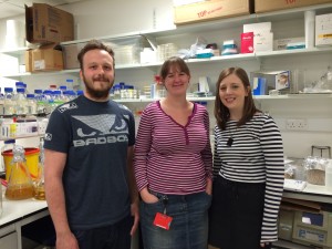
Researchers Max Temple, Lis Lowe and Fiona Cuskin of the Gilbert lab.

Researchers Max Temple, Lis Lowe and Fiona Cuskin of the Gilbert lab.

Yeast is commonly in our diet in the form of some of our favourite things: bread, beer, wine and fermented food products.  So it may be dry January but if your willpower fails and you succumb to an alcoholic beverage, then make sure it’s an unfiltered one with plenty of yeast!

This research in the Gilbert lab was funded by the Wellcome Trust and the European Research Council.

Links:

Nature article: http://www.nature.com/nature/journal/v517/n7533/full/nature13995.html

Press release: http://www.ncl.ac.uk/press.office/press.release/item/beer-and-bread-yeast-eating-bacteria-aid-human-health

Gilbert Lab: http://www.ncl.ac.uk/camb/staff/profile/harry.gilbert


Discovery at the Discovery Museum

Aside


Great Hall

Great Hall

Ready to replicate the success of last year’s Away Day, it was en masse outing time for ICaMB again! Time to find out who all the new faces are, and to find out exactly what that person you have a ‘hello and a nod in the corridor’ relationship with actually does at the bench all day. This year we headed to the Great Hall of the Discovery Museum. Despite the leaking roof caused by the downpour outside and the sometimes dodgy acoustics the day was still a success.

Serious faces, this is science

Serious faces, this is science

ICaMB is a fast paced, constantly evolving institute. Everybody is busy with their own research, making a break and a get-together once in a while a vital part of reminding ourselves of the vast range of expertise and diverse set of interests beavering away in our labs and offices. The answer to that tricky problem or that elusive technique is quite possibly just a few yards away.

But also fun!

But also fun!

With that in mind, this year’s Away Day felt particularly important as we welcomed 8 new academic groups to ICaMB from CAV (Campus for Aging and Vitality) as well new IRES fellows and a list of other recent recruits. Drs Victor Korolchuk and Gabi Saretzki from the CAV both spoke at the away day about their interests in neurodegenerative diseases and the role of oxidative stress in the ageing process.

As ever the day was kicked off by the Institute director, Bob (Professor Robert Lightowlers), who gave us a taster of ICaMB’s growth and success stories over the past year. Without breaking into the tune of that well known Christmas song; 7 Vacation Studentships, 6 BBSRC awards, 5 MRC awards, 2 Wolfson awards, 2 Senior Investigator awards and 1 Henry Dale ………. Not to mention all the promotions, outstanding research papers and commercial contracts = 1 happy Bob.

ICaMBAwayDay1(2)

ICaMBAwayDay3


A cell-tastic morning then ensued: the completely dispensable nature of bacterial cell walls (Professor Jeff Errington); the role of NF-kB in the pathogenesis of lymphoma (Dr Jill Hunter); and the cell death independent functions of inhibitors of apoptosis (new IRES recruit Dr Niall Kenneth). The session was wrapped up by Dr Paula Salgado summarising 3.5 years of structural C. difficile research in 15min. Some feat Paula!

Of course just as last year, an absolute highlight of the day were the six, animated, three minute thesis presentations by our brave PhD students ……..  Soon to be followed by the look of horror on several Professorial faces when it was suggested by PAN!C that at next year’s Away Day we have a session of 3 minute PI pitches! We can’t ignore the demands of our PhD students now can we? And congratulations to Mandeep Atwal from the Cowell/Austin lab who against steep competition was awarded the prize for best three minute thesis.

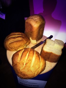
The possibilities of alginate bread?

The possibilities of alginate bread?

A spot of oxidative stress and the evolution of peroxidases by Dr Alison Day, and some lunch completed the morning’s discovery. Though half an hour later and Dr Peter Chater had us all wishing we’d had an alginate packed lunch (and a go with the model gut!). Perhaps the Pearson lab can cater next year’s event? If it’s good enough for the One Show it’s good enough for the ICaMB Away Day.

A major focus of the Away Day is not just to learn about the breadth of exciting research carried out in our institute, but also to learn all about the very latest techniques and expertise ripe for exploitation. This year the focal point of new techniques came from Dr Alex Laude and the Bio-Imaging facility, with some beautiful images and super resolution microscopy techniques, which again left a number of the audience wanting a turn!

P1000375An afternoon transcribing and translating with Dr Danny Castro-Roa; learning about how the crucial nature of cell polarity means we really don’t mix up our arse from our elbow (thank you new IRES recruit Dr Josana Rodriguez); and last but by no means least, how on earth all that DNA manages to faithfully copy and repackage itself time after time from yet another new recruit, Professor Jonathan Higgins.

This completes our diverse and entertaining line-up, just leaving enough time for complementary wine, and the amusement as speakers and audience alike embarrass themselves at the ICaMB quiz (and I hear also in the pub afterwards).

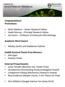
More ICaMB winners! Doctoral Thesis Prize Success.

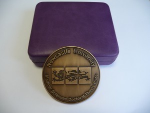
‘The Faculty of Medical Sciences Doctoral Thesis Prize is a mark of recognition of an outstanding level of achievement by the end of a research doctorate. Prizes are awarded biannually on a very limited basis following nomination by thesis examiners.’ Dr Tim Cheek, Post Graduate Tutor

Doctoral Bling!

Doctoral Bling!

Prizes were first awarded in 2009 and included two ICaMB students, Holly Anderson and Monika Olahova. This was followed in 2011 by David Adams and in 2012 by Graham Scholefield. However, 2013, was an absolute triumph, with three out of only five potential Faculty Prizes being bestowed on theses submitted by ICaMB students. Dr Andrew Foster from Professor Nigel Robinson’s Lab (currently a post-doc in the Robinson lab in Durham), Dr Fiona Cuskin from Professor Harry Gilbert’s lab (currently a post-doc in the Gilbert lab) and Dr Kristoffer Winther from Professor Kenn Gerdes lab (currently a post-doc in Gerdes lab). With their new roles keeping them busy, our 3 winners only just managed to get together recently to be presented with their medals by the Dean of Post Graduate studies. Andrew and Fiona tell us about their past and present research.

Dr Andrew Foster

Dr Andrew Foster

Abstract by Dr Andrew Foster. Achieving metal selectivity is often more difficult than one might first imagine as the inherent chemical properties of metals often mean that a metalloprotein will preferentially select an incorrect metal over a correct one.

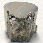
My PhD studies involved understanding metal selectivity among a group of proteins called metal sensors. These metal sensing, transcriptional regulators control the expression of genes of metal homeostasis and therefore influence the metallation of other proteins within the cell. I characterised a novel nickel sensor InrS and showed for the first time how metal selectivity could correlate with relative metal affinity across a class of proteins. The nickel sensor InrS has a tighter nickel affinity than the other sensors within the cell, thus InrS responds to nickel activating

Nickel

Nickel

a nickel efflux gene so that the buffered nickel concentration within the cell does not rise high enough to mis-populate the sensors of other metals.

During my PhD studies our lab moved from Newcastle to Durham University but I remained registered at Newcastle. This move was obviously very disruptive but at the same time made me more focussed and determined to make a success of the work in spite of the disruption.

Busy Andrew

Busy Andrew

I am currently working with Professor Nigel Robinson at Durham University. My current work seeks to understand how the affinity of a metal sensor relates to the available concentration of the sensed element within the cell. Our model system involves the nickel sensor I discovered, InrS, and nickel supply to hydrogenase, a nickel enzyme capable of hydrogen production. Metal supply to enzymes will be a key biotechnological challenge as we seek to utilise microbial factories for the production of fuel and other useful products.

Dr Fiona Cuskin.

Dr Fiona Cuskin.

Abstract by Dr Fiona Cuskin. The use of complex carbohydrates in the food industry is wide and varied; a few examples include the use of polysaccharides and oligosaccharides as gelling agents, emulsifiers and fat replacements. Small oligosaccharides are being increasingly used as prebiotics for the vast array of “friendly” bacteria in the gut of both humans and animals. The addition of small fructose oligosaccharides by the food industry into yoghurts, amongst other foods, has been shown to promote a healthy gut flora, which in turn has a positive effect on the host gut health and immune system.

Having been in the lab for just a month my supervisor abandoned me and moved to America. Not to worry I tracked him down and moved there too for a few months. The subject of my PhD was to investigate how bacteria use enzymes called glycoside hydrolases to breakdown complex carbohydrates for utilisation. Part of this was to characterise a glycoside hydrolase that degraded the fructose containing polysaccharide, levan.This glycoside hydrolase contained two

Happy gut!

Happy gut?

modules, the catalytic module and non-catalytic carbohydrate-binding module (CBM). CBMs are usually attached to enzymes that catalyse the breakdown of recalcitrant insoluble substrates to help target the catalytic module to the right carbohydrate. However, the CBM characterised in my PhD bound soluble fructan polysaccharides and potentiated the activity of the catalytic module ~100 fold. This work adds valuable knowledge to how bacteria breakdown complex polysaccharides. This knowledge can be exploited to better inform the use of prebiotics and to also choose enzymes that are efficient for the production of small oligosaccharides from polysaccharides.

We are very proud of our current winners. Who will be in the next batch of Doctoral Thesis Prize winners, adding to a growing list of ICaMB winners?


Skinny Seaweed

Matt

Obesity has reached epidemic proportions globally, with at least 2.8 million people dying each year as a result of being overweight or obese (WHO). Dr Matthew Wilcox from Professor Jeff Pearson’s lab tells us about his research into the slimming powers of brown seaweed. A simple and sustainable food additive. 

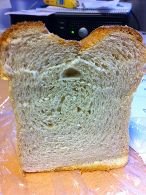
“Seaweed bread” by Dr Matt Wilcox

Seaweed bread

Seaweed bread

Over 40% of the calories in your diet can come from fat (mainly triacylglycerol) and you digest and absorb between 95 and 100% of all the fat that you eat!  There is one enzyme (pancreatic lipase) that accounts for 80% of all fat digestion and if the activity of this enzyme can be reduced then we will absorb less fat from our diet.

The Pearson lab has shown that alginate (a dietary fibre extracted from brown seaweed) can reduce the activity of pancreatic lipase by up to 80%.  If you don’t digest fat you can’t absorb it.

Times pictureLipase is a recognised target as a treatment for obesity. In 2010 the pharmaceutical drug orlistat (Xenical) which acts on this enzyme accounted for 98% of all UK prescriptions for the treatment of obesity. The remaining 2% was for Sibutramine which has since been withdrawn. We are therefore heavily reliant upon one drug which can have some rather unpleasant side effects (brown trouser syndrome) greatly reducing patient compliance. So even though it’s the only pharmaceutical drug available, not everyone who is prescribed it will actually take it!

Tasteless alginate is already found in a number of foods (E400-405), salad dressings, ice-creams ..... even beer.

Tasteless alginate is already found in a number of foods (E400-405), salad dressings, ice-creams ….. even beer.

Thankfully there is still hope (and clean trousers) since some, if not all of the side effects of orlistat can be eliminated if it is taken with a high fibre product. Luckily, it just so happens that alginate is not only a lipase inhibitor but is also a dietary fibre itself, and to top it all off makes great bread!

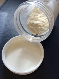
Most commercial alginates come from seaweed, hence the seaweed bread.  The extraction process is relatively simple and you end up with a dry white (ish) powder, great to use in products with flour. There is no ‘seaweed’ taste (although seaweed bread is actually quite nice) and in blinded tests people actually prefer the alginate bread over standard white bread.  Likely to be due to the retention of moisture so it’s like eating fresh bread, even if it’s a day or so old

Seaweed culture and harvesting is big business around the world from the Shetlands to Shanghai and can even be seen from space. The extracted alginates are already used in the food industry, just not necessarily the right ones or in the right concentrations. The majority we test in the lab are from Norwegian or Scottish seaweeds.

The  latest technology in seaweed harvesting.

The latest technology in seaweed harvesting.

The alginate project in the lab started nearly eight years ago with simple colourimetric assays. These initial experiments showed that certain seaweed alginates can inhibit lipase. From this we developed a model of the upper gastrointestinal tract to test the alginates in a system as close to a human as possible without being human. Our model gut has now been used to show that alginate is released from the bread (amongst other things) and has also featured in a BBC3 program. We have just finished the first proof of concept trial in humans, so there will be more on this story to follow.

The Pearson Lab line-up

The Pearson Lab line-up

The big fat truth is that fat in food is often vital for the enjoyment, try eating cream crackers without butter (3 cracker challenge).  It’s hard!  So, if we can still get the enjoyment but not all the calories from our food then we can significantly reduce the amount of calories we take in.  It’s not just a reduction in the absorption of fat in the food that contains the alginate; it’s the entire meal that you eat.  Not that you should eat loads of burgers but if we could put alginate into burger buns then you would reduce the amount of fat absorbed from the burger, the cheese, the fries and the ridiculously oversized milkshake that you have with it.

Low fat lunch?

Low fat lunch?

Perhaps that’s the best way to end a blog with the thought of a ridiculously oversized milkshake and also by thanking the BBSRC for all their funding and support!

Matt’s PhD was a BBSRC CASE studentship sponsored by Technostics

This work has been patented

Full Links

The patent: http://worldwide.espacenet.com/publicationDetails/biblio?DB=worldwide.espacenet.com&II=1&ND=3&adjacent=true&locale=en_EP&FT=D&date=20121129&CC=US&NR=2012302521A1&KC=A1

Technostics: http://www.technostics.com/

Matthew Wilcox: http://www.ncl.ac.uk/camb/staff/profile/matthew.wilcox

Jeff Pearson: http://www.ncl.ac.uk/camb/staff/profile/jeffrey.pearson

ICaMB Research Update: Zenkin lab Science paper

Congratulations to ICaMB and CBCB researchers Soren Nielsen, Yulia Yezenkova and Nikolay Zenkin and  who have a paper published in the prestigious journal Science today.

Nikolay Zenkin: RNA Polymerase researcher

There are three RNA Polymerases in Eukaryotic cells and although much attention focuses on the role of RNA Polymerase II, since it transcribes mRNAs from protein encoding genes, it is easy to forget that the majority of transcription in the cell is accomplished by RNA Polymerase III.  RNA Polymerase III (or Pol III as it is commonly known) is responsible for the synthesis of ribosomal 5S rRNA, tRNA and other small RNAs.


Soren’s paper, entitled “Mechanism of Eukaryotic RNA polymerase III Transcription” termination solves a long-standing mystery in the field of how transcriptional termination by RNA polymerase III takes place. Their study reveals an elegant scenario, in which co-transcriptional folding of highly-structured RNA polymerase III transcripts causes termination at the end of their genes. This mechanism ensures proper folding of the structural/catalytic RNAs synthesized by RNA polymerase III prior to RNA release. Analogies with bacterial termination suggest that this fundamental mechanism may have emerged before divergence of bacteria and eukaryotes.

Here is the Science Editor’s summary of the paper

It is as important to terminate any biological process as it is to start it. Transcription, copying information encoded in genes into RNA, requires accurate and timely termination. Nielsen et al. (p. 1577) present a mechanism for transcription termination by RNA polymerase III, the enzyme that synthesizes the majority of RNA molecules in eukaryotes. In this scenario, the folding of the RNA as it is transcribed by polymerase into a highly structured transcript causes termination at the end of its synthesis. This mechanism may serve as a control of proper folding of structural or catalytic RNAs synthesized by RNA polymerase III. Comparison with other organisms suggests that this mechanism emerged before divergence of bacteria and eukaryotes.

And here is the abstract of their paper

Gene expression in organisms involves many factors and is tightly controlled. Although much is known about the initial phase of transcription by RNA polymerase III (Pol III), the enzyme that synthesizes the majority of RNA molecules in eukaryotic cells, termination is poorly understood. Here, we show that the extensive structure of Pol III–synthesized transcripts dictates the release of elongation complexes at the end of genes. The poly-T termination signal, which does not cause termination in itself, causes catalytic inactivation and backtracking of Pol III, thus committing the enzyme to termination and transporting it to the nearest RNA secondary structure, which facilitates Pol III release. Similarity between termination mechanisms of Pol III and bacterial RNA polymerase suggests that hairpin-dependent termination may date back to the common ancestor of multisubunit RNA polymerases.

Links

Link to the Science paper: http://www.sciencemag.org/content/340/6140/1577.abstract

Nikolay Zenkin laboratory home page: http://www.ncl.ac.uk/camb/staff/profile/nikolay.zenkin#tab_research

Centre for Bacterial Cell Biology: http://www.ncl.ac.uk/cbcb/

Science magazine: http://www.sciencemag.org/

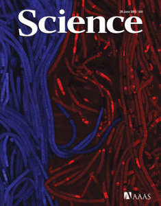
Link to the Science cover: http://www.sciencemag.org/content/340/6140.cover-expansion