More ICaMB winners! Doctoral Thesis Prize Success.

‘The Faculty of Medical Sciences Doctoral Thesis Prize is a mark of recognition of an outstanding level of achievement by the end of a research doctorate. Prizes are awarded biannually on a very limited basis following nomination by thesis examiners.’ Dr Tim Cheek, Post Graduate Tutor
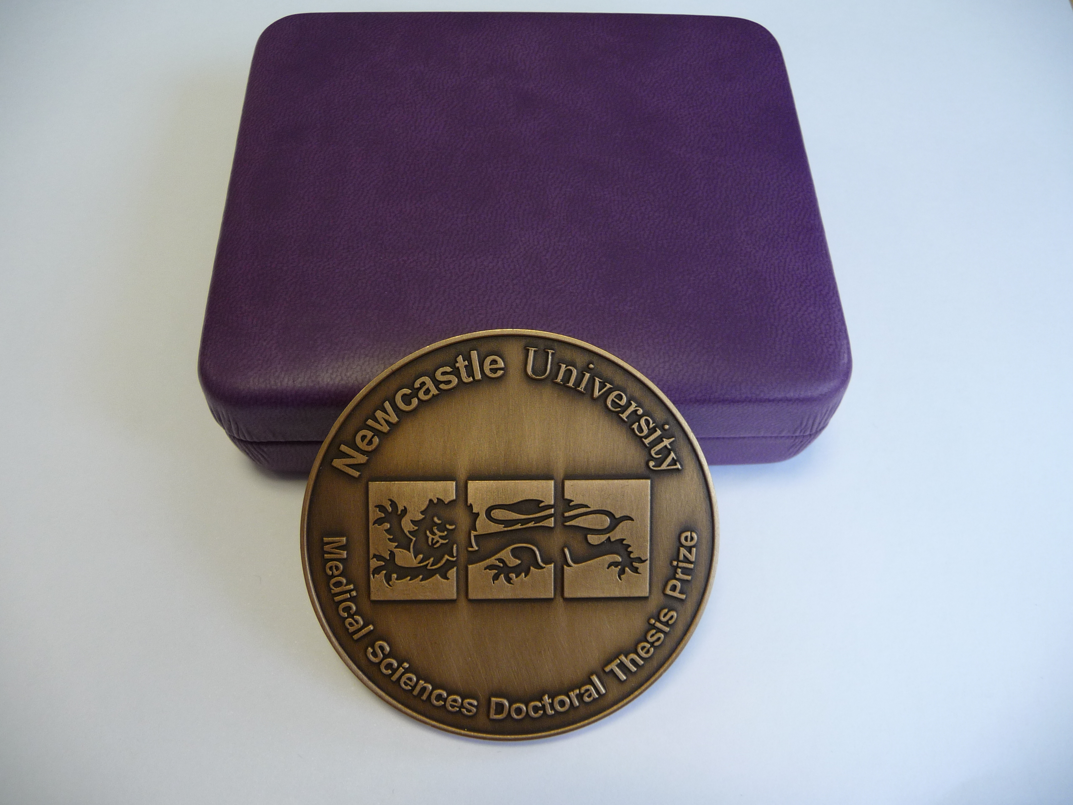
Doctoral Bling!

Doctoral Bling!

Prizes were first awarded in 2009 and included two ICaMB students, Holly Anderson and Monika Olahova. This was followed in 2011 by David Adams and in 2012 by Graham Scholefield. However, 2013, was an absolute triumph, with three out of only five potential Faculty Prizes being bestowed on theses submitted by ICaMB students. Dr Andrew Foster from Professor Nigel Robinson’s Lab (currently a post-doc in the Robinson lab in Durham), Dr Fiona Cuskin from Professor Harry Gilbert’s lab (currently a post-doc in the Gilbert lab) and Dr Kristoffer Winther from Professor Kenn Gerdes lab (currently a post-doc in Gerdes lab). With their new roles keeping them busy, our 3 winners only just managed to get together recently to be presented with their medals by the Dean of Post Graduate studies. Andrew and Fiona tell us about their past and present research.
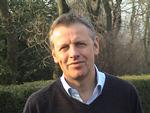
Dr Andrew Foster

Dr Andrew Foster

Abstract by Dr Andrew Foster. Achieving metal selectivity is often more difficult than one might first imagine as the inherent chemical properties of metals often mean that a metalloprotein will preferentially select an incorrect metal over a correct one.

My PhD studies involved understanding metal selectivity among a group of proteins called metal sensors. These metal sensing, transcriptional regulators control the expression of genes of metal homeostasis and therefore influence the metallation of other proteins within the cell. I characterised a novel nickel sensor InrS and showed for the first time how metal selectivity could correlate with relative metal affinity across a class of proteins. The nickel sensor InrS has a tighter nickel affinity than the other sensors within the cell, thus InrS responds to nickel activating
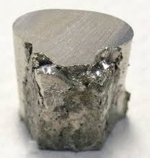
Nickel

Nickel

a nickel efflux gene so that the buffered nickel concentration within the cell does not rise high enough to mis-populate the sensors of other metals.

During my PhD studies our lab moved from Newcastle to Durham University but I remained registered at Newcastle. This move was obviously very disruptive but at the same time made me more focussed and determined to make a success of the work in spite of the disruption.
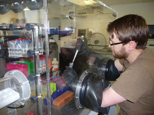
Busy Andrew

Busy Andrew

I am currently working with Professor Nigel Robinson at Durham University. My current work seeks to understand how the affinity of a metal sensor relates to the available concentration of the sensed element within the cell. Our model system involves the nickel sensor I discovered, InrS, and nickel supply to hydrogenase, a nickel enzyme capable of hydrogen production. Metal supply to enzymes will be a key biotechnological challenge as we seek to utilise microbial factories for the production of fuel and other useful products.

Dr Fiona Cuskin.

Dr Fiona Cuskin.

Abstract by Dr Fiona Cuskin. The use of complex carbohydrates in the food industry is wide and varied; a few examples include the use of polysaccharides and oligosaccharides as gelling agents, emulsifiers and fat replacements. Small oligosaccharides are being increasingly used as prebiotics for the vast array of “friendly” bacteria in the gut of both humans and animals. The addition of small fructose oligosaccharides by the food industry into yoghurts, amongst other foods, has been shown to promote a healthy gut flora, which in turn has a positive effect on the host gut health and immune system.

Having been in the lab for just a month my supervisor abandoned me and moved to America. Not to worry I tracked him down and moved there too for a few months. The subject of my PhD was to investigate how bacteria use enzymes called glycoside hydrolases to breakdown complex carbohydrates for utilisation. Part of this was to characterise a glycoside hydrolase that degraded the fructose containing polysaccharide, levan.This glycoside hydrolase contained two
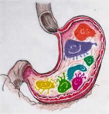
Happy gut!

Happy gut?

modules, the catalytic module and non-catalytic carbohydrate-binding module (CBM). CBMs are usually attached to enzymes that catalyse the breakdown of recalcitrant insoluble substrates to help target the catalytic module to the right carbohydrate. However, the CBM characterised in my PhD bound soluble fructan polysaccharides and potentiated the activity of the catalytic module ~100 fold. This work adds valuable knowledge to how bacteria breakdown complex polysaccharides. This knowledge can be exploited to better inform the use of prebiotics and to also choose enzymes that are efficient for the production of small oligosaccharides from polysaccharides.

We are very proud of our current winners. Who will be in the next batch of Doctoral Thesis Prize winners, adding to a growing list of ICaMB winners?


Deep Impact

Alginate bread on a pedestal under show-biz lights

Alginate bread on a pedestal under show-biz lights.


Another excellent post by ICaMB’s Dr Matthew Wilcox as the fame of alginate spreads and seaweed bread goes on tour!

Well, I was kindly invited along to the BBSRC Fostering Impact awards ceremony in London a couple of weeks ago and although I wasn’t up for anything, Newcastle University were.

Fostering impact is a scheme run by the BBSRC to capture the economic and social impact of research funded by them.  There are three competitions that fit the fostering impact scheme; ‘innovator of the year’, ‘activating impact’ and ‘excellence with impact’.  The first is for an individual researcher, the second is for the knowledge exchange teams and the final award is for research organisations (runs from 2013 – 2015).

Outside eventInside eventThe knowledge exchange and commercialisation team at Newcastle University has changed substantially over the past couple of years.  What was once a centralised Business Development Directorate has now become the Research Enterprise Service, comprised of three teams, each embedded in one of the Faculties.  Each Institute or School now has their own dedicated business development manager (BDM), with ICAMB’s BDM being Laura Rush (who is very nice).  They are now much easier to contact, whether it’s just a quick question or the drafting of patents.

Home baking

Home baking practice.

Newcastle’s application for the Activating Impact award was submitted back in October 2013 and used the wonderful research done by the beautiful people of ICAMB as its basis.  In January the RES team found out that they had successfully made it to the final five (from 18) and through to the grand final in London.  Newcastle was up against the knowledge exchange and commercialisation teams from King’s college London, Queen Mary University of London, University College London and University of Aberdeen. One of the requirements of the competition was to bring along someone to the final who had worked with RES, a ‘user’ (according to the BBSRC).  They also wanted an iconic object?!  Alginate bread it was then.  Back in the kitchen I went. How many loaves would I need to feed the people there? Two should do it, right?

Martin and Laura on the train gearing up for competition.

Martin and Laura on the train gearing up for competition.

In London Martin Cox presented the case for Newcastle in front of a panel of scientists, business types and other technology transferers, assembled by the BBSRC. Demonstrating what Newcastle does well, how BDM’s have been embedded into each institute and also what they would do with the money if we won (£100k).  He also described the additional internal funds that are available to help activate impact.  FMS has two funds available; the first is to help with data collection for translational grant applications, the second is to support further claims in patent applications.  The two internal grants can both potentially support a post doc salary for three months, plus consumables.

Dengue fever carrying mosquito

Dengue fever carrying mosquito.

The awards ceremony combined the ‘innovator of the year’ and ‘activating impact’.  I got a glitzy stand for my bread and also had the chance to look around the other pretty amazing stuff that was on display, like Dr Luke Alphey’s work. Luke ended up being named both social innovator and overall innovator of the year for his work on the genetic control of pests, including the dengue fever carrying mosquito.

I even got to meet the new (ish) CEO of the BBSRC, Professor Jackie Hunter, who was definitely not snapped stuffing free stuff into her bag!

BBSRC's CEO Jackie Hunter enjoying the exhibition

On the right BBSRC’s CEO Professor Jackie Hunter enjoying the exhibition.

Unfortunately Newcastle did not win, but being down to the last five of the competition is brilliant and should give confidence to ICAMB scientists that when help is required in achieving impact (social or economic), we have a great team to help.

Queen Mary University of London, whose entry was also being supported by a previous BBSRC Enterprise Fellow and King’s College London, ended up being joint winners each scooping £100k.

Perhaps if a few more of the world leading researchers in ICaMB engaged with the Enterprise team, they might not have to only take some eejit and his bread to the competition next year and increase NU’s chance of winning!

Pruning the Tree of Life

Dr Tom WIlliams

Anyone who has studied biology has seen an image of the tree of life in the text books.  Most of us think of this as being set in stone, one of the rock solid foundations on which evolutionary biology is built.  However, all is not quite as settled as it seems.  Recently, a Nature article from the laboratory of ICaMB’s Professor Martin Embley challenges the traditional three domain structure of the root of life.  Here, first author on the paper, Dr Tom Williams, tells us the story.

By Dr Tom Williams

Our modern understanding of the tree of life began in 1977 when Carl Woese and his colleagues discovered the Archaea, a group of prokaryotes originally isolated from extremely hot or salty environments. Although Archaea looked indistinguishable from Bacteria under the microscope, their gene sequences were at least as different to those of Bacteria as from the eukaryotes – the group of organisms, including fungi, animals and plants, whose cells contain a mitochondrion and a nucleus. According to these analyses, living cells should be classified into three main groups: Bacteria, Archaea and eukaryotes – rather than the two (prokaryotes and eukaryotes) that had previously been established based on cell structure. In 1990, Woese and his colleagues published another seminal paper in which they argued for this “three domains” classification. This three-domains tree has become an iconic image in biology, and is often found in the popular science literature, as well as many textbooks – you’ve probably seen it before. Here it is from a 1997 review by Norman Pace:

The traditional 3 domain Tree of Life. From: A molecular view of diversity and the biosphere. Pace NR Science (1997) 276: 734-740


Professor Martin Embley

This was certainly the tree of life that I was familiar with, first as an undergrad and later as a Ph.D. student at Trinity College Dublin. So I was surprised and very intrigued when a certain Martin Embley came to talk at an Irish bioinformatics meeting, claiming that support for the three-domains tree was not as strong as you might expect. New work from his lab instead favoured the “eocyte tree”, in which the eukaryotes (or, at least, some of their genes) actually evolved from within the Archaea. If true, this tree would imply that there were originally only two types of cells – Bacteria and Archaea – and that the eukaryotes (i.e., us!) originated later in a partnership between the two primary domains.

The new model of the Tree of Life proposed by the Embley lab

Fast-forward a couple of years, and I was thinking about where I wanted to do my postdoc. I remembered Martin not only from that talk, but also from some interesting work (2nd link) he had done on a group of parasitic fungi called Microsporidia. I joined his group and began working on microsporidians, but I was still very interested in the tree of life and the origin of eukaryotes. In the meantime, DNA sequencing technology had been improving, and microbial ecologists were beginning to publish genomes from new groups of Archaea that could not be grown in the lab, and so had never been studied before. One of the really exciting findings from these studies was that some Archaea contained genes that looked very similar to fundamental components of our own cells, such as actin and tubulin – two proteins that help to define the microscopic “skeleton” of eukaryotic cells. When we added these new genomes to our analyses, we found even stronger support for the eocyte tree; those findings were reported last year in Proceedings B. At about the same time, a number of other researchers were reporting something similar: as our view of archaeal biodiversity increased, support for the three-domains tree was on the wane. Given the prominent position of the three-domains tree in the literature, and the importance of this question for understanding early life on Earth, we decided to write a review summarizing these recent developments in the field – it came out in Nature this week, and it’s the reason for this blog post!

As we delved back into the 30 years of literature on the molecular tree of life, one of the most interesting discoveries for me was a seam of eocyte literature that I hadn’t been aware of previously. Although many analyses over the past three decades have recovered the three-domains tree, and it appears in all the textbooks, the literature has actually never been unanimous in its support. Nonetheless, it is only in the last five years or so that support for the eocyte hypothesis has reached critical mass, perhaps due to improvements in our statistical methods and, more recently, sampling of archaeal biodiversity.

The Embley lab: Back row, left-to-right: Kacper Sendra, Martin Embley, Tom Williams, Robert Hirt. Front row: Shaojun Long, Ekaterina Kozhevnikova, Andrew Watson, Paul Dean, Maxine Geggie,Alina Goldberg-Cavalleri, Sirintra Nakjang.

Of course, our latest work is almost certainly not going to be the last word on the relationship between eukaryotes and other cells. Our methods are getting better – in part thanks to the statisticians we are collaborating with here in Newcastle – but there is much room for improvement, and so much about the microbial world that we still have to discover. Still, if the eocyte tree is correct – and it appears to be the best-supported tree on the current evidence – then that has important implications for how we understand early life on Earth and the origin of our own cells. For one thing, it rules out the eukaryotes as a primordial cellular lineage, as old as the Bacteria and Archaea. Instead, it suggests that the Bacteria and Archaea were established and diversifying on Earth before the origin of eukaryotes, resurrecting the concept of an “Age of Prokaryotes” on the early Earth. Of course, when you think about the phenomenal number of Bacteria and Archaea that live in your own body, never mind the wider environment, you might well argue that it never ended…

This work was supported by a Marie Curie postdoctoral fellowship to Tom Williams. Martin Embley acknowledges support from the European Research Council Advanced Investigator Programme and the Wellcome Trust.

Links

The Nature Article: http://www.nature.com/nature/journal/v504/n7479/full/nature12779.html?WT.ec_id=NATURE-20131212

The Proceedings B paper: http://rspb.royalsocietypublishing.org/content/279/1749/4870

The Embley lab website: http://research.ncl.ac.uk/microbial_eukaryotes/

Microsporidia papers: http://www.nature.com/nature/journal/v452/n7187/full/nature06606.html

http://www.nature.com/nature/journal/v453/n7194/full/nature06903.html

Breaking news: Mitochondria replacement therapy given green light

Breaking news by Prof Bob Lightowlers

Professor Dame Sally Davies, the Governments Chief Medical Officer, has today given support to a change in the law that will allow the form of mitochondrial replacement therapy that is being pioneered here in Newcastle, has we mentioned before.

Its great news for any prospective mother with mitochondrial DNA disease who is concerned about transmitting the disease to their children.

Most media is covering this – BBC, Guardian, Telegraph – but if you are still confused, we thought it would be good to recap the essential points:

Electron micrograph of a cell (coloured blue) revealing part of the mitochondrial structure (orange) within. The entire length of the mitochondrion is about 5 micrometres.

– Mitochondria are crucial structures found in all cells in our body and they take our common foodstuffs such as fats and sugars and turn them into energy. They have their own genetic element, mitochondrial (mt) DNA. Much smaller than our chromosomes, mtDNA is essential for energy production.

– In 1988, scientists in the UK and US recognised that certain diseases were caused by mutations in mtDNA with the main disorders related to your muscle tissue and the brain.

– It is estimated that at least 1:10,000 people suffer from disorders associated with defects in mtDNA – that’s more than 6,000 people in the UK.

– Mitochondrial DNA is only transmitted to babies by their mothers. Unfortunately, as you inherit your mothers mitochondria, diseases caused by mtDNA mutations are inadvertently transmitted from the mother.

– Doug Turnbull, Director of the Wellcome Trust Centre for Mitochondrial Research (WTCMR) and I have wondered since the 1990s whether it would be possible to prevent the transmission of the faulty mtDNA from the mothers to their children by transferring the nucleous from an egg of the mother carrying these mtDNA mutations into a healthy egg whose nucleous had been removed, and then fertilise it

– Professor Mary Herbert, Doug and a group of us from the Mitochondrial Research Group were able to show that such a swap could be performed without any or very low levels of the defective mtDNA being transferred. Importantly, there was also no defect detectable in the reconstituted cells.

In August 2012, the government asked the Human Fertility and Embryological Authority (HFEA) to find out what the general public thought of the procedure.  The results were collated  and the Human Fertility and Embryological Authority made a recommendation to Government. There was an overall support for the new technology with only 10% being fairly or strongly against the concept of mitochondrial gene replacement

To understand all the details and find out the whole story, see my post a while ago here.

The Wellcome Trust also has a very nice video explaining the science behind the news story:


Links

Wellcome Trust Centre for Mitochondrial Research http://www.newcastle-mitochondria.com/
Human Fertility and Embryological Authority (HFEA) http://www.hfea.gov.uk/index.html
HFEA mitochondria puclib consultation 2012 http://www.hfea.gov.uk/6896.html


Science Minister visits Centre for Bacterial Cell Biology


by Dr Heath Murray 

On June 27 the RH David Willetts MP, Minister for Universities and Science, visited the Centre for Bacterial Cell Biology (CBCB) to hear about how research on bacteria can lead to: development of novel antibiotics, design of synthetic biological systems, and even understanding the origins of life on earth. Dr Heath Murray (CBCB & ICaMB) tells us more about this visit.

Mr. Willetts was given a guided tour of the new Baddiley-Clark building by the director of the CBCB, Prof Jeff Errington.

Jeff (left) outlines CBCB research to David Willetts (right), with Heath (middle back) paying close attention

Jeff discussed why he left Oxford University after 25 years to start the CBCB at Newcastle, the first Centre of its kind in the UK to provide a world-class facility in which to carry out fundamental research on bacterial cells. During the tour Jeff highlighted how the localised network of international researchers at the CBCB, working on biological problems in model bacterial organisms provides an unparalleled setting in which to exchange ideas and to benefit from related advances in microbial cell biology. While walking around Jeff noted how the open plan of the Baddiley-Clark building promoted interactions amongst the various research groups, thereby creating a uniquely stimulating environment for the scientists that work there.

This was a very fruitful visit with interesting discussions, as highlighted by Jeff: “I was impressed at how quickly the Minister picked up the key biological points we wanted to make, such as about how our work impacts on thinking about the origins of life!

An image similar to those seen by David Willetts showing severe DNA segregation defect in a mutant Bacillus subtilis strain, observed using epifluorescent microscopy. (DNA: blue; origin of replication: green, cell membrane: red)


I then demonstrated the bespoke microscopes available within the CBCB to the Minister, highlighting how the small size of bacterial cells (only a few micrometers) makes microscopic analysis technically challenging and how the CBCB is utilizing state-of-the-art super-resolution microscopes to overcome this difficulty. I also explained how researchers use genetic engineering to fuse their “proteins of interest” to the Green Fluorescent Protein (GFP) from the jellyfish Aequorea victoria, thus creating tools to visualize the localization of proteins or nucleic acids within living bacterial cells using fluorescence microscopy.


Heath explains the potential applications of the research to the Science Minister


The Minister was keen to see the live demonstration of our fluorescent microscope and seemed amazed by how clearly the organization of the bacterial chromosomes was immediately apparent. He quickly appreciated that interfering with this process might have application in the development of new antibiotics.


We were all left with the clear feeling that Mr. Willetts enjoyed hearing about the science taking place within the CBCB and how this fundamental research provides insights crucial for the discovery and development of new antibiotics, as well as providing solutions to a wide range of industrial and environmental problems. “It was an interesting meeting – very reassuring to hear that the Minister is keen to make sure that Government continues to invest in Blue-Skies Research”, Jeff concluded.


You don’t always want what your mother gives you! – can we prevent mitochondrial disease?


By Professor Robert Lightowlers

In 1988, scientists in the UK and US recognised that certain diseases were caused by mutations in mtDNA . Over the following 20 years, mtDNA defects have been shown to cause a range of debilitating diseases many affecting different parts of the body. However, the main disorders relate to your muscle tissue and the brain.

Human muscle fibres stained for mitochondrial function. As can be seen in B, some of the fibres show no activity. This is because these fibres have high levels of mutated mitochondrial DNA.

It is estimated that at least 1:10,000 people suffer from disorders associated with defects in Mitochondrial DNA (mtDNA) – that’s more than 6,000 people in the UK. Even so, it is only recently that the importance of mitochondrial diseases have hit the general media.

Many of you will have seen the debate on correcting mitochondrial diseases in the newspapers (for example, see the Guardian, Telegraph) and on television recently, but not be aware of the central role that Newcastle researchers have played in making this exciting, or to some, controversial, new therapy closer to becoming a reality.  Here, Bob Lightowlers ICAMB Director and senior member of the Wellcome Trust Centre for Mitochondrial Research (WTCMR) reflects on the role mitochondrial research in Newcastle has played in this process over the last 20 years and tells us some of the story behind the headlines.

What are Mitochondria?

Electron micrograph of a cell (coloured blue) revealing part of the mitochondrial structure (orange) within. The entire length of the mitochondrion is about 5 micrometres.


These crucial structures found in all the trillions of cells in our body have many essential functions. One very important role they play is to take our common foodstuffs such as fats and sugars and turn them into energy for our body’s to function.

A single human cell showing the nucleus (green), the mitochondrial network (red) and the mitochondrial DNA within the network (yellow)


One surprising element of these structures is that they contain their own genetic element, mitochondrial (mt) DNA. Much smaller than our chromosomes, mtDNA is essential for energy production.


OK, so this is important, but why have mitochondria and mtDNA begun to work their way into the common conversation of the nation?

Answer: Our mothers!

What has this got to do with our mothers ? Mitochondrial DNA is only transmitted to babies by their mothers. This is different to all our other DNA where copies are made and transmitted from both parents. Unfortunately, as you inherit your mothers mitochondria, diseases caused by mtDNA mutations are inadvertently transmitted from the mother.

How does this relate to Newcastle based Mitochondrial Research?

My colleague Doug Turnbull, a neurologist here in Newcastle (and Director of the WTCMR) and I have been intrigued by these mtDNA mutations since it first became clear that they could cause disease. Back in the early ‘90’s, we discussed whether some day it would be possible to try and prevent the transmission of the faulty mtDNA from the mothers to their children. Of course, at that stage, it was just wishful thinking. As the Mitochondrial Research Group (MRG) began to grow and mature in Newcastle, we often returned to one question:

What if the nucleus from the diseased egg could be transferred to a healthy egg whose nucleus had been removed, in essence leaving all the affected mtDNA behind ?

If it was indeed possible, this reconstituted egg could be fertilised and implanted back into the mother by standard techniques used routinely in fertility clinics throughout the world. We also would consider when would such a technique be most efficient: before or after fertilisation of the egg? On paper both options looked possible, but there are many complications.

Technical Concept: achieving the switch of nuclei without some of the faulty mtDNA being inadvertently taken along for the ride.

Towards the end of the 90’s, scientists working in Canada were able to show that the level of mtDNA inadvertently transferred when the nucleus was switched into a recipient cell lacking a nucleus, was low. This was a promising result, but it led to two central questions:

•   Could this be repeated with human cells?

•   Was this technique morally and ethically acceptable to everyone?

The ethical debate: Debate raged as to whether this technique would constitute genetic manipulation of humans, which of course would be illegal. Further, it was not possible to perform these types of reconstruction experiments in man, as using viable human fertilised cells for research was also, understandably, illegal.

Professor Mary Herbert working at the nearby Human Fertility Centre came up with an intriguing proposal. She explained that unfortunately, during the standard process of in vitro fertilisation, many eggs became incorrectly fertilised. These eggs are unable to grow correctly and have to be discarded. One way of determining whether it would be possible to swap mtDNA in humans, she suggested, was to use these incorrectly fertilised eggs. As this procedure would still require the manipulation of fertilised human eggs, a licence would need to be applied for from the Human Fertilisation and Embryological Authority (HFEA). Following lengthy and extensive debate, including members of the research team being called to the House of Commons, a licence was eventually awarded in 2005.  Five years later, with the essential help of colleagues in the Fertility Centre, Mary, Doug and a group of us from the MRG were able to show that such a swap could be performed without any or very low levels of the defective mtDNA being transferred. Importantly, there was also no defect detectable in the reconstituted cells . In 2011, this very promising result, along with many other important contributions made by the Newcastle MRG to understanding mitochondrial biology in health and disease was recognised by the Wellcome Trust who funded the establishment of a new Research Centre in Newcastle, the Wellcome Trust Centre for Mitochondrial Research.

Getting acceptance of the technique

It was important to know whether the people of the UK agreed that such reconstitution technology was ethically acceptable. In August 2012, the government asked the Human Fertility and Embryological Authority (HFEA) to find out what the general public thought of the procedure .  The results were collated last month and the Human Fertility and Embryological Authority made a recommendation to Government. There was an overall support for the new technology with only 10% being fairly or strongly against the concept of mitochondrial gene replacement ) This is an endorsement of the method but there is still a long way to go before the technique can be performed in the clinic.

Its amazing to think how far this concept has come in 20 years. Perhaps in another 20 years we may be able to look back and celebrate how this dream has helped to provide a realistic method to help prevent the transmission of a debilitating disease for many couples.


Wellcome Trust Centre for Mitochondrial Research http://www.newcastle-mitochondria.com/
Human Fertility and Embryological Authority (HFEA) http://www.hfea.gov.uk/index.html
HFEA mitochondria puclib consultation 2012 http://www.hfea.gov.uk/6896.html

Postgraduate Newsbite I


One of the aims of our blog is to make it a forum to share what is happening, so this week we focus on upcoming events important for our postgraduate students.

Firstly, the Postgraduate Research Symposium is taking place on Monday, 25th. This is a unique opportunity for all of us to hear more about the exciting research being carried out by our students. It’s also a great occasion for the students, a chance for them to share their work with the whole Institute and practice those ever important presentation skills.

So please come and show your support on Monday 25th March, 9.30 to 4.30, in Lecture Theatre D. Don’t forget you also get a chance to meet the students over lunch, provided in the Boardroom. We will also cover details of the day in our Blog next week – so come back then to find out more!

ICaMB’s Postgraduate association, PAN!C, start their academic events this afternoon at 4pm, in the Baddiley-Clark seminar room.  ICamBlog regular Jeff Errington will be giving a “Careers talk” where he will discuss his own experiences in academia and in setting up two spin out companies from his research. MRes, PhD students and Postdocs are all welcome.

Also, next Wednesday, 27th March, PAN!C social events continue with a pub quiz at Mr Lynch in Jesmond at 8.30pm.

We will soon hear about their plans for future events, so watch this space for more news from PAN!C.

Jeff Errington Wins Novartis Medal

Congratulations to ICaMB‘s Professor Jeff Errington, who has just been awarded the prestigious Biochemical Society Novartis Medal and Prize for 2014.

The Novartis Medal and Prize is an annual award that recognises contributions to the development of any branch of biochemistry. Work leading up to the award must be carried out in the UK but is open to all nationalities. A list of previous winners can be seen here. A full list of the 2014 prize winners is here.

Jeff moved to Newcastle in 2005 where he became Director of ICaMB before standing down in 2012.  He founded the Centre for Bacterial Cell Biology (CBCB) in 2007, which has since become a world leading centre for microbial research with a strong interest in aiding the discovery of the next generation of antibiotics.  Jeff’s lab is best known for its ground breaking work on the bacterial cell cycle and cell morphogenesis.  On the ICaMB blog we recently highlighted his Cell paper on L-form bacteria and insights into primordial cell division.

This is the video where Jeff describes the work in this paper

Early work from the Errington lab led to the formation in 1998 of  Prolysis, an antibacterial drug discovery company, which was recently acquired by an international anti-infectives company Biota Pharmaceuticals Inc.  More recently, a Newcastle University spin out company Demuris Ltd has been established to exploit drug screening opportunities emerging from the Errington lab.

In ICaMB, we are all delighted that Jeff’s work has received this recognition.  Above all it proves that you can achieve success in science while still being a a great guy who loves his beer and football.


Exploding bacteria for science!!!

As the Chief Medical Officer, Professor Dame Sally Davies, highlights today “danger posed by growing resistance to antibiotics should be ranked along with terrorism on a list of threats to the nation”. Professor Dame Sally Davies said diseases are evolving faster than the drugs that exist to treat them and antibiotic resistance is “a ticking time bomb“.

This is a subject of great interest to scientists in ICaMB, particularly the Centre for Bacterial Cell Biology, which brings together a world-class group of scientists researching bacterial physiology and the host response to bacterial infections. A major focus of this research involves:

  • Exploring alternative targets for antibiotic development
  • Understanding how antibiotics attack bacterial cells
  • Investigating how bacteria overcome such an attack

Tonight (March 11th 2013), work from the group of Kenn Gerdes and Etienne Maisonneuve, a post-doc in his group, will be featured on the BBC programme “Bang Goes the Theory” in an episode about Antibiotics.

Kenn and Etienne’s research focuses on persister cells, bacterial cells that can tolerate and survive attack by antibiotics.  Importantly, ALL bacteria analysed so far generate “persister cells” and understanding this is key to understanding how bacteria avoid antibiotic attack. “Bang Goes the Theory” will show a movie showing how these persister cells are identified in a bacterial population.

Penicillin inhibits synthesis of the bacterial cell wall, causing the cell to explode (or ‘lyse’) due to the high pressure inside the cell.  This is why penicillin and similar drugs are very effective in curing infections caused by penicillin-sensitive bacteria. In the movie, see how the cells suddenly explode when penicillin is added but notice how one cell, the persister cells (darker cells not exploding on the left panel) are surviving.

These persister cells evade killing by antibiotics because they grow extremely slowly. Persisters are proposed to be one explanation for infection relapses or chronic infections so Kenn and Ethienne’s work is extremely important for understanding how we should use antibiotics.

Microfluidic chamber used to make the movie

To do this work, Etienne used state-of-the-art technology – microfluidics – to follow the growth of individual bacterial cells under a microscope. These devices are smaller than a penny coin and the chambers where the bacteria are grown can be less than 1 mm across. This technique allows us to grow bacteria in one condition but, at a flip of a switch, change it and watch the response, as seen in the movie.

Year 9 student working on one of CBCB's microscopes


CBCB academics have used the ability to explode Escherichia coli to explore the what, when and how of antibiotics with Year 9 school students as part of the University engagement program Leading Edge.

With these students, we have developed a protocol to allow them to observe E. coli in the act of exploding after adding penicillin.

Exploding E. coli. Taken by Seaton Burn Community College Year 9 students

Persistence is not Resistance: It is important to understand the difference between these two terms. Antibiotic resistant and sensitive bacteria are able to generate persister cells, that are not effected by antibiotic attack. Antibiotic Resistance is a trait acquired by the whole population.

The Scientific Specifics: Over the last few years, several scientific breakthroughs made by the Gerdes group have, for the first time, given insight into how bacteria control the switch to slow growth and persistence. Persister cells can survive penicillin because the bacteria hibernate for a period, during which they don’t synthesize their cell wall.  They can then “wake up” when the antibiotic treatment is over, causing a new infection. In young and healthy people this is usually not a problem, because the rare non-growing bacteria are removed by the immune system. However, elderly individuals or those with a weakened immune system, it is often not efficient enough to permit clearance of the rare bacteria that survive the treatment, allowing the infection to “break out”.

The Gerdes group has shown that a certain class of gene that inhibits cell growth are turned on in one cell per 10,000. These discoveries open avenues to generate novel antibiotics and treatment regimes in the future. However, before that, their group is investigating if similar mechanisms allow pathogenic bacteria, such as Mycobacterium tuberculosis, to evade killing by antibiotics.


Institute of Cell and Molecular Biosciences
The Centre For Bacterial Cell Biology
Professor Kenn Gerdes
Bang Goes the Theory
Leading Edge

Bulging bacteria and the origins of life

Jeff (left), Romain (centre) and Yoshikazu (right), the team of researchers behind these exciting discoveries


In a paper published this week in CellJeff Errington’s team in ICaMB, have discovered new insights into the origin of life on Earth.


Jeff and his team share their results

Bacteria were the first organisms to appear on planet earth. Almost all modern bacteria have a tough protective shell called a cell wall. The structure of the wall and the mechanisms used by cells to manufacture it are conserved, suggesting that the wall was invented right at the beginning of bacterial evolution, and, therefore, when the first true cells emerged.

Production of cell wall is carefully regulated by complex machineries that allow the cell to enlarge and then divide in a controlled manner, all the time maintaining the integrity of the wall intact.

Despite its importance, it seems that many modern bacteria can survive cell wall loss under certain very special conditions, such as when they are treated with certain antibiotics that interfere with its production, like penicillin. Not only that, but a few years ago my lab showed that these “L-form” cells (named after the Lister Institute in London where they were first described) no longer need the complex mechanisms normally needed for bacterial growth and division. Instead, they grow by extrusion of irregular tubes or blebs of cytoplasm, that pinch off into daughter cells.

Our team – me, Yoshikazu KawaiRomain Mercier – has been working on this problem for some time. “Studying L-form biology is a real technical challenge, and this work could not have succeeded without the strong collaboration established between us“, says Romain. As Yoshikazu explains: “we developed a very simple genetic system to isolate mutations enabling L-form development from non-viable protoplasts.

We are excited because we think we have now solved the mystery of how L-forms grow and divide. Our latest results, published in Cell, show that the mechanism is remarkably simple: it requires only that cells make excess amounts of membrane – the thin porous layer that acts as the outer boundary of all cells, including our own.

Increasing the membrane surface area beyond the amount needed to contain the cytoplasm causes the cell to buckle and distort. Eventually, this leads to pinching off of membrane bags that are ill formed but nonetheless viable “baby” cells.

Time-lapse photography representing the division of B. subtilis without cell wall (L-form). The images were obtained using light microscopy. Scale bar: 3 μm

At first, we thought this mechanism was too simple to be true, we changed our minds when we were alerted to amazing experiments being done by several groups working on the origins of life, particularly Jack Szostak at Harvard, Saša Svetina in Ljubljana and Peter Walde in Zurich. These groups have been wondering how primitive cells could have arranged to grow and divide efficiently without spilling all of their contents. They recently found that simple membrane bags, called “vesicles”, can be induced to grow and reproduce into multiple smaller vesicles, in the test tube, just by increasing their surface area.

So, in explaining how the bizarre L-form bacteria manage to survive the loss of their beloved cell wall, we think we may now also have glimpsed how the first primitive cells could have duplicated themselves at the dawn of life on earth.

Jeff Errington 
Director of the Centre for Bacterial Cell Biology


Cell paper: http://www.cell.com/abstract/S0092-8674(13)00135-9
Cell website: http://www.cell.com/home see PaperFlick
Newcastle University Press Release:http://www.ncl.ac.uk/press.office/press.release/item/how-did-early-primordial-cells-evolve#.US-chen77jQ

Soapbox Science guest blogpost: http://www.blogs.nature.com/soapboxscience/2013/02/28/social-media-from-an-institutional-perspective-why-are-we-on-there

ICaMB website: http://www.ncl.ac.uk/camb/
Facebook: https://www.facebook.com/pages/ICaMB-Newcastle/416200498466481
Twitter: https://twitter.com/ICaMB_NCL
YouTube: http://www.youtube.com/channel/UCSuZgA6URiXTUoHq1tMe-PQ