How a motile cytoskeleton drives bacterial cell division

seamusIn a recent issue of Science, the discovery of a key mechanism for bacterial cell division was reported. This work was carried out by Dr Seamus Holden’s lab (ICaMB) in collaboration with Professor Cees Dekker (TU Delft), Professor Yves Brun (Indiana University), Professor Mike VanNieuwenhze (Indiana University) and Professor Ethan Garner (Harvard University). Here, Seamus tells us about this discovery and what its implications could be for antimicrobial research.

Bacterial cell division is a lovely mechanistic problem in biology: how do the simplest living organisms build a crosswall at mid cell, against very high outwards pressure (think of a racing bike tyre), without bursting?  A ring of protein filaments forms around the future division site, and enzymes associated with this ring build a new crosswall that cleaves the bacteria in half. But what has remained completely mysterious is how these proteins work together as a single nanoscale machine to cut the bacterial balloon skin (cell wall) in two.

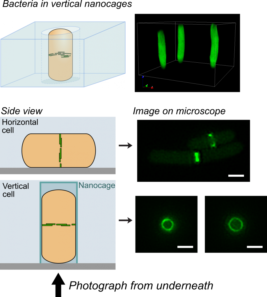
Cytoskeletal proteins FtsZ in live bacteria imaged in vertical nanocages

Cytoskeletal proteins FtsZ in live bacteria imaged in vertical nanocages

Working together with collaborators in Delft, Indiana and Harvard, we tracked the organization and motion of key division proteins as they build the dividing crosswall, and the organization of the newly built crosswall itself. We began by examining the motion of FtsZ, a cytoskeletal filament that is required for cell division – cytokinesis – in bacteria and is related to the tubulin cytoskeletal protein found in eukaryotic cells. Using high-resolution microscopy techniques, we found that FtsZ filaments move around the division site, traveling around the division ring. We imaged the motion of individual cell wall synthesis enzymes, and saw that the synthesis enzymes ride on FtsZ filaments, building new cell wall as they travel along the division site. This causes the cell wall to be synthesized in discrete sites that travel around the division site during cytokinesis, a process which we were able to observe directly by using dyes that label the bacterial cell wall. Using a variety of experimental techniques, we were able to speed up or slow down how fast FtsZ rotated around the cell. Strikingly, we found that the speed of FtsZ filament motion determines how fast the cell can divide. When FtsZ moves more rapidly, cell wall is produced more quickly, and cytokinesis happens faster. This shows that the motion of FtsZ is the critical overall controller of cell division.

One challenge that we faced was trying to look at the division proteins in actively dividing cells. At the earliest stages of division, it was possible to image division protein organization because the proteins in the partially assembled ring are sparsely distributed. However, a new strategy was required to measure how the dense protein network of actively dividing cells was organized. Normally, bacteria are immobilized flat on a microscope slide, and imaged from underneath, but unfortunately this places the division ring side-on, obscuring the motion and organization of division proteins. To solve this problem, we used nanofabrication technology, originally developed to manufacture computer chips, to create tiny gel nanocages to trap bacteria in an upright position.

Bacteria trapped in vertical nanocages

Bacteria trapped in vertical nanocages

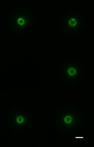
By trapping individual bacteria upright, we were able to rotate the cell division ring so that it was fully visible on our high resolution microscope. This revealed the dynamic motion of FtsZ filaments as they travel around the entire division site:

Together, these results revealed the basic mechanistic principles of bacterial cell division: that the building of the division crosswall is orchestrated by moving cytoskeletal filaments.  Previously, the cytoskeleton was thought to serve as a static scaffold, recruiting other molecules and perhaps exerting some force to divide the cell. This new work demonstrates that all the components of cell division are in constant, controlled motion around the division site, driven by the fundamental dynamics of the cytoskeleton.

In the longer term, this study could open up novel antibiotic targets. Based on the discovery that the treadmilling motion of the bacterial cytoskeleton is critical for division, it may be possible to develop new drugs that specifically inhibit this motion, similar to how the chemotherapy drug taxol suppresses the motion of the cytoskeleton in cancer cells.

==================================

Links

Explanatory animation: https://youtu.be/6dq2_gqKPfU (Animation credit TU Delft / Scixel)

Nanocage Video: nanocage-movie-2.

Science report: http://science.sciencemag.org/content/355/6326/739

Press release: http://bit.ly/2kwsnJf

Any excuse to drink red wine

By Elisabeth Lowe

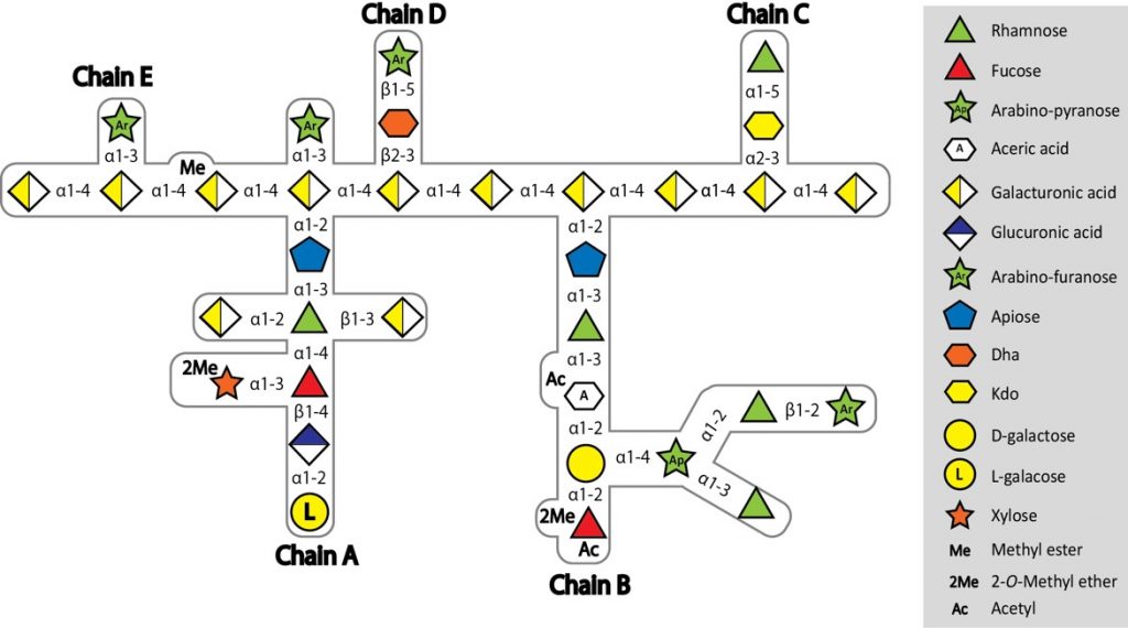
I’m writing this blog from an interesting perspective – though a member of the lab I’m not involved in the project, except through the (exhaustive!) discussions at coffee time with Harry Gilbert and the rest of the authors on the paper. This work, published today in Nature, describes the way in which one particular species of human gut bacteria is able to degrade a complex plant polysaccharide. Why is this new, you might well ask? Well, after being talked to death on the project, so much so I’d be glad never to hear a word about it again, I’ve actually come to appreciate what a great piece of work it is. The Bolam and Gilbert labs have published quite a bit on plant polysaccharide degradation over the years, but this one is a little different, because the polysaccharide involved – Rhamnogalacturonan-II (RGII) – is thought to be the most complex glycan found in nature. It contains ~13 different sugars linked by 21 different linkages, and all but one can be broken by a single bacterial species, Bacteroides thetaiotaomicron (Bt).

Complex structure of RG-II from red wine

Complex structure of RG-II from red wine

Wade Abbott, a post-doc in Harry’s lab when he was at the Complex Carbohydrate Research Centre, in Athens, Georgia, which is incidentally where the structure of RG-II was determined in the first place by Alan Darvil and Malcolm O’Neill in the 1980s, initiated this project. We knew which loci were upregulated during growth on RG-II from earlier work by myself, Dave Bolam and Eric Martens, so Wade set about cloning and expressing the 30 genes in these loci, and looking for activity with a PhD student, Jeff Xhang. It was a tough job, and little progress was made. When Harry came back to the UK he decided he absolutely had to crack it, and put two post-docs onto the project, Art Rogowski and Didier Ndeh. Though as the project has progressed, more and more people from the lab were sucked in, including the post-docs Alan Cartmell and Aurore Labourel, and Harry’s last PhD student, Ana Sofia Luis. All worked incredibly hard, assisted by Arnaud Baslé from SBL, and between them discovered the activities of 26 enzymes, including seven entirely new glycoside hydrolase families, three new activities, and seven crystal structures, with all the active site mutations, and ligand soaks that go along with a new crystal structure.

Working out the enzyme activities on a polysaccharide as complex as RG-II poses some problems, in that you can’t really buy it, so you have to make it. RGII is concentrated in red wine, so Harry put 150 litres of Asda’s not so finest wine on Dave Bolam’s credit card (don’t ask) and with Art and Didier, took it to Prozomix in Haltwhistle, to use their industrial concentrators.

Concentrating wine

Starting to concentrate 150 L of wine

As you can see, this did not end well.

It's all gone horribly wrong

It’s all gone horribly wrong


Art then turned to apple concentrate and managed to purify some through an intensive multiple two-week process. Making or buying oligosaccharides is also extremely difficult, so Didier devised a way of killing two birds with one stone – identifying which unknown open reading frames (ORFs) were enzymes, and making oligos at the same time. By deleting the gene of interest, RG-II degradation (which is mainly exo-acting) was halted at the point at which that enzyme acted. Taking the supernatant from these strains grown on RG-II and purifying the sugars released by the bacterium yielded partial breakdown products which could be identified by mass spectrometry with tremendous help from Joe Gray, and then used as a substrate for the enzyme in question.

Getting the data in a format suitable for the tight space restrictions imposed by Nature was almost as challenging as doing the work. Given that Harry is not very artistic, and certainly has no idea how to use Adobe Illustrator, he was fortunate to have three talented artists Ana, Art and Aurore, who ended up doing all the figures. They are now referred to as the art department. This project was definitely a labour of love (for Harry, if no-one else!) and by carefully characterising the breakdown of this polysaccharide, one linkage at a time, has actually revealed new features of the structure of this critical plant polysaccharide. Finding and characterising the vast repertoire of enzymes produced by gut microbes is not only interesting in its own right, but can provide tools to understand complex glycan structure. It’s a fantastic achievement, and I pity the poor lab member who has to try and bake the RG-II cake for the celebrations this week!


Link to paper


Origins of life in Newcastle

ICaMB’s Prof Jeff Errington organised and hosted an impromptu symposium on the origins of life at the Centre for Bacterial Cell Biology (CBCB) on Wednesday 18th January 2017. About 80 people attended, hearing 11 talks from a mixture of Newcastle and international speakers, including a number of guests who had travelled over from Japan for the meeting. The program was arranged more or less in “chronological” order, starting with the origins of the solar system 4-6 billion years ago, and ending (still almost 2 billion years in the past) with the emergence of the eukaryotes. The meeting sparked several very lively discussions, perhaps reflecting the difficulty of doing standard hypothetico-deductive experiments on the topic, in the absence of time travel technology! Nevertheless, the day was a great success and is likely to lead to new international collaborations and funding opportunities.

By Jeff Errington

My emerging interest in the subject has two origins. First, through ageing and trying to find a reason for existence before existence disappears! Second, the lab’s work on L-form bacteria (see Box), which has attracted much interest from the origins of life scientific community.

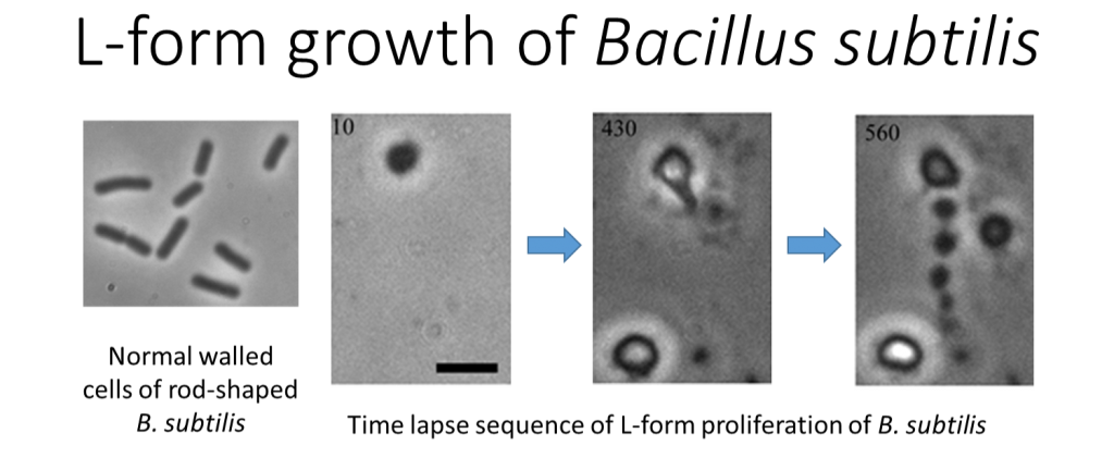
L-form bacteria use a seemingly primitve mechanism of replication.

L-form bacteria use a seemingly primitive mechanism of replication. L-forms are cell wall deficient bacteria, which turn out to replicate by a slightly bizarre, seemingly haphazard mechanism involving membrane blebbing and tubulation. The process provides a model for how primitive life may have proliferated billions of years ago, before the invention of the cell wall.


The latest findings have led to a number of fascinating new scientific contacts, and about a year ago, Prof Shige Maruyama, who heads a major Japanese research institute dedicated to origins of life work called the Earth-Life Science Institute (ELSI), made contact, proposing discussion around possible collaborations. After a series of small meetings in Newcastle and my visit to Tokyo, momentum began to emerge, culminating with the proposal for a major workshop in Newcastle, with half a dozen or so ELSI members planning to attend.

Prof Shige Maruyama, ELSI, Tokyo, Japan

Prof Shige Maruyama, ELSI, Tokyo, Japan

As discussions developed, I identified various experts in Newcastle with complementary expertise and interests in the general area, and the idea for a full blown symposium took shape. There was even time to identify a top class international “guest” speaker, Prof Bill Martin from Dusseldorf, who came over at short notice to give the concluding talk.

This is not the place to go through each talk in detail. However, from my perspective, what I hope people took away from the meeting would have included the following general points.

First, the problem is amazingly multidisciplinary, with important contributions from astrophysicists, geochemists, organic chemists, microbiologists (structure/function, metabolism and physiology) and evolutionary bioinformaticians. Second, we still have a very hazy understanding of many of the early events in the earth’s planetary history, e.g. when did the water arrive and how much? Third, it is clear that microbes were responsible for huge changes in planetary chemistry, particularly oxygenation but also that planetary composition must have reciprocally influenced microbial evolution.

Prof Bill Martin, Dusseldorf, Germany

Prof Bill Martin, Dusseldorf, Germany

The day concluded with a very nice dinner at the Jesmond Dene House Hotel, supported by Newcastle University and hosted by Pro-Vice Chancellor Prof Nick Wright. I’m sure that the original owner of the house, Lord Armstrong, would have approved of the day (for example, I gather that his company won the contract to build ships for the Japanese Navy 120 or so years ago). I’m also sure that as a Fellow of the Royal Society (elected in 1843) he would have been acquainted with Charles Darwin and perhaps they too had interesting conversations about the origins of life in their own time frame.

Beer, Bread and Bacteria


Prof. Harry Gilbert

Prof. Harry Gilbert

The diverse organisms that live in our gut, collectively termed the gut microbiota, can have a major impact on our health. A new paper from Prof. Harry Gilbert’s lab in ICaMB, published today in Nature, shows that one member of our gut microflora uses a ’selfish’ mechanism to degrade a component of the cell wall of the yeast in our diet.

By Drs. Elisabeth Lowe and Fiona Cuskin.

Have you over-indulged this Christmas? One too many ales, or too many turkey sandwiches?

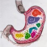
Beer: full of yeast.

Beer: full of yeast.

The good news is that, while your waist line may be expanding and you may be feeling a little the worse for wear, the organisms of your gut microbiota are thriving!

Some of the bacteria in the human gut, called Bacteroides, can break down the complex carbohydrates in our diet into simple sugars, which they use for energy.

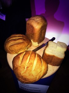
Bread: full of yeast


Our work, published in Nature today, shows that one of these bacteria can also break down some of the carbohydrate cell wall of yeasts, called mannan, a polymer of mannose. This includes mannan from Saccharomyces cerevisiae (Baker’s yeast), and the pathogenic gut fungus, Candida albicans. The current understanding of polysaccharide digestion by gut bacteria suggests a cooperative environment, in which break-down products are shared between different members of the microbiota. However our data shows that this species (Bacteroides thetaiotaomicron) is ‘selfish’ when it degrades mannan, and doesn’t share any of the digestion products with other bacteria.

Turkey: er... full of yeast?

Turkey: er… full of yeast?

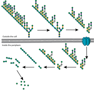
To achieve this selfish degradation, the large and multiply-branched mannan structure undergoes only very limited degradation in the extracellular space (see figure). The resulting processed mannan fragments are then imported into the Bacteroides periplasmic space, where they are further degraded by a suite of glycoside hydrolase enzymes to yield the sugar mannose. Achieving complete breakdown within the periplasm prevents  the mannose from being shared with competing bacteria.

This selfish approach to mannan utilisation could offer the opportunity to design bespoke prebiotics targeted to a specific bacterial population within the gut. Food products containing yeast mannan would be expected to promote growth of B. thetaiotamicron, whereas other complex polysaccharides may specifically promote other members of the microbiota.

Complex polysaccharide mannan is sequentially degraded by a 'selfish' mechanism to yield periplasmic mannose.

Complex polysaccharide mannan is sequentially degraded by a ‘selfish’ mechanism to yield periplasmic mannose.

A number of inflammatory bowel diseases such as Crohn’s disease are associated with intolerance to yeast, and particularly cell wall polysaccharides, and have also been linked to low levels of Bacteroides species in the gut. Our work provides a potential mechanism for how Bacteroides might be able to influence the effect of yeast on patients with Crohn’s disease, and in fact a drug named Thetanix (which is a live formulation of Bacteroides thetaiotaomicron) has been licensed for treatment of paediatric Crohn’s disease in the USA.

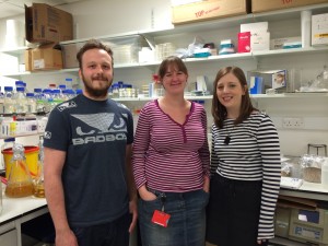
Researchers Max Temple, Lis Lowe and Fiona Cuskin of the Gilbert lab.

Researchers Max Temple, Lis Lowe and Fiona Cuskin of the Gilbert lab.

Yeast is commonly in our diet in the form of some of our favourite things: bread, beer, wine and fermented food products.  So it may be dry January but if your willpower fails and you succumb to an alcoholic beverage, then make sure it’s an unfiltered one with plenty of yeast!

This research in the Gilbert lab was funded by the Wellcome Trust and the European Research Council.

Links:

Nature article: http://www.nature.com/nature/journal/v517/n7533/full/nature13995.html

Press release: http://www.ncl.ac.uk/press.office/press.release/item/beer-and-bread-yeast-eating-bacteria-aid-human-health

Gilbert Lab: http://www.ncl.ac.uk/camb/staff/profile/harry.gilbert


A CBCB Cell-ebration

Heath MurrayKevin WaldronEarlier this month, the Centre for Bacterial Cell Biology held its inaugural Symposium. Here, the CBCB’s Heath Murray and Kevin Waldron tell us about what happened at the event.

One of the aspects of ICaMB that makes it a unique institute is the Centre for Bacterial Cell Biology (CBCB), a group of researchers who are focused on understanding fundamental biological questions using bacteria as model organisms. The CBCB was founded by Professor Jeff Errington FRS and is the world’s first major research centre with a focus on bacterial cell biology. Since its inception, CBCB has relocated to a purpose-built £30 million facility in the Baddiley-Clark Building, and has grown to include more than 20 different research groups. In a relatively short time, CBCB members have made outstanding contributions to our understanding of numerous aspects of fundamental cellular processes in a wide range of bacteria.

Prof Kenn Gerdes from the CBCB discusses how bacteria can form dormant variants that evade the immune defence response.

Prof Kenn Gerdes from the CBCB discusses how bacteria can form dormant variants that evade the immune defence response.

In order to recognise the success and the breadth of science being generated in the Centre, we recently held the inaugural CBCB Symposium on July 9-10. More than 120 members of the CBCB community participated in the two-day event, underscoring the critical mass of researchers at Newcastle University working within the field. This excellent turnout certainly contributed to the overall success of the event.

Research themes covered by talks from group leaders in the CBCB included sporulation, infection, persistence, biofilms, metabolism, motility, and morphogenesis. We also heard about the emerging subject of synthetic biology, where bacterial organisms will be programmed much like computers to perform discrete biological tasks.The CBCB Symposium was highlighted by inspirational talks from three distinguished external scientists, Jan Löwe (Laboratory of Molecular Biology, Cambridge), Mervyn Bibb (John Innes Centre, Norwich), and Simon Foster (Department of Molecular Biology and Biotechnology, Sheffield).

Prof Simon Foster explains how the superbug Staphylococcus aureus grows and divides.

Prof Simon Foster explains how the superbug Staphylococcus aureus grows and divides.

Professor Löwe discussed his work using a range of biochemical and structural approaches to analyse the bacterial cell division and morphogenesis machinery. Professor Bibb explained how his lab utilises a combination of next generation DNA sequencing and bioinformatics with classical genetic analysis to discover novel antibiotics. Professor Foster showed how studies on the fundamental aspects of bacterial cell biology can be harnessed to better understand host-pathogen interactions that can eventually be translated into vaccine development, with his focus on the ‘super bug’ Staphylococcus aureus..

Participants hold discussions over dinner and drinks following the Symposium.

Participants hold discussions over dinner and drinks following the Symposium.

At the end of the Symposium participants gathered together for dinner and drinks in the informal setting of the Forum. This provided an interactive end to the event that allowed researchers throughout the CBCB to meet one another, discuss the amazing science, and develop connections.

More ICaMB winners! Doctoral Thesis Prize Success.

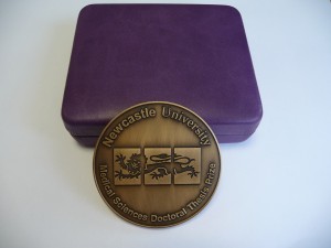
‘The Faculty of Medical Sciences Doctoral Thesis Prize is a mark of recognition of an outstanding level of achievement by the end of a research doctorate. Prizes are awarded biannually on a very limited basis following nomination by thesis examiners.’ Dr Tim Cheek, Post Graduate Tutor

Doctoral Bling!

Doctoral Bling!

Prizes were first awarded in 2009 and included two ICaMB students, Holly Anderson and Monika Olahova. This was followed in 2011 by David Adams and in 2012 by Graham Scholefield. However, 2013, was an absolute triumph, with three out of only five potential Faculty Prizes being bestowed on theses submitted by ICaMB students. Dr Andrew Foster from Professor Nigel Robinson’s Lab (currently a post-doc in the Robinson lab in Durham), Dr Fiona Cuskin from Professor Harry Gilbert’s lab (currently a post-doc in the Gilbert lab) and Dr Kristoffer Winther from Professor Kenn Gerdes lab (currently a post-doc in Gerdes lab). With their new roles keeping them busy, our 3 winners only just managed to get together recently to be presented with their medals by the Dean of Post Graduate studies. Andrew and Fiona tell us about their past and present research.

Dr Andrew Foster

Dr Andrew Foster

Abstract by Dr Andrew Foster. Achieving metal selectivity is often more difficult than one might first imagine as the inherent chemical properties of metals often mean that a metalloprotein will preferentially select an incorrect metal over a correct one.

My PhD studies involved understanding metal selectivity among a group of proteins called metal sensors. These metal sensing, transcriptional regulators control the expression of genes of metal homeostasis and therefore influence the metallation of other proteins within the cell. I characterised a novel nickel sensor InrS and showed for the first time how metal selectivity could correlate with relative metal affinity across a class of proteins. The nickel sensor InrS has a tighter nickel affinity than the other sensors within the cell, thus InrS responds to nickel activating

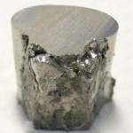
Nickel

Nickel

a nickel efflux gene so that the buffered nickel concentration within the cell does not rise high enough to mis-populate the sensors of other metals.

During my PhD studies our lab moved from Newcastle to Durham University but I remained registered at Newcastle. This move was obviously very disruptive but at the same time made me more focussed and determined to make a success of the work in spite of the disruption.

Busy Andrew

Busy Andrew

I am currently working with Professor Nigel Robinson at Durham University. My current work seeks to understand how the affinity of a metal sensor relates to the available concentration of the sensed element within the cell. Our model system involves the nickel sensor I discovered, InrS, and nickel supply to hydrogenase, a nickel enzyme capable of hydrogen production. Metal supply to enzymes will be a key biotechnological challenge as we seek to utilise microbial factories for the production of fuel and other useful products.

Dr Fiona Cuskin.

Dr Fiona Cuskin.

Abstract by Dr Fiona Cuskin. The use of complex carbohydrates in the food industry is wide and varied; a few examples include the use of polysaccharides and oligosaccharides as gelling agents, emulsifiers and fat replacements. Small oligosaccharides are being increasingly used as prebiotics for the vast array of “friendly” bacteria in the gut of both humans and animals. The addition of small fructose oligosaccharides by the food industry into yoghurts, amongst other foods, has been shown to promote a healthy gut flora, which in turn has a positive effect on the host gut health and immune system.

Having been in the lab for just a month my supervisor abandoned me and moved to America. Not to worry I tracked him down and moved there too for a few months. The subject of my PhD was to investigate how bacteria use enzymes called glycoside hydrolases to breakdown complex carbohydrates for utilisation. Part of this was to characterise a glycoside hydrolase that degraded the fructose containing polysaccharide, levan.This glycoside hydrolase contained two

Happy gut!

Happy gut?

modules, the catalytic module and non-catalytic carbohydrate-binding module (CBM). CBMs are usually attached to enzymes that catalyse the breakdown of recalcitrant insoluble substrates to help target the catalytic module to the right carbohydrate. However, the CBM characterised in my PhD bound soluble fructan polysaccharides and potentiated the activity of the catalytic module ~100 fold. This work adds valuable knowledge to how bacteria breakdown complex polysaccharides. This knowledge can be exploited to better inform the use of prebiotics and to also choose enzymes that are efficient for the production of small oligosaccharides from polysaccharides.

We are very proud of our current winners. Who will be in the next batch of Doctoral Thesis Prize winners, adding to a growing list of ICaMB winners?


ECRs at ICaMB: Copying the blueprint of life – Understanding DNA replication


Heath MurrayIn the latest of our series focussing on the Early Career Researchers (ECRs) in ICaMB, we feature Dr Heath Murray.  After completing undergraduate studies at the University of California, Los Angeles and then obtaining his Ph.D. from the University of Wisconsin-Madison, Heath came to the UK to join the lab of Prof Jeff Errington in Oxford. From there he re-located to ICaMB, and in 2009 was awarded a Royal Society University Research Fellowship. Here, Heath describes his research into the mechanisms of DNA replication, and explains why he became interested in this field.

By Dr Heath Murray

Hello, my name is Heath Murray and I’m a Royal Society University Research Fellow in ICaMB’s Centre for Bacterial Cell Biology (CBCB) studying DNA replication. DNA is one of the most important molecules required for life because it encodes the information, or the blueprint, used to build a cell (i.e. the most basic unit of an organism). In order for a cell to create new cells it must synthesize an exact copy of its DNA, an extraordinary process when you consider that the genomes of most cells contain millions of individual DNA subunits!

Bacillus subtilis is a useful model system as it proliferates rapidly and is amenable to genetic, cell biological, biochemical, and structural analyses

Bacillus subtilis is a useful model system as it proliferates rapidly and is amenable to genetic, cell biological, biochemical, and structural analyses

Bacteria are ideal model systems to study this fundamental process because they are much less complex than human cells (e.g. all of their DNA is encoded by a single chromosome, whereas humans have 23), and this allows us to understand how they work at the greatest possible level of detail.

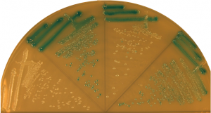
I was introduced to bacteria when I was an undergraduate student and the effect was transformative. My mentors taught me how to add a specific gene (a DNA sequence) to a bacterial cell, and if it worked properly then the bacteria would turn blue!

Bacterial colonies turn blue if they contain a gene that degrades specific sugars.

Bacterial colonies turn blue if they contain a gene that degrades specific sugars.

That basic genetic experiment was one of the coolest things I had ever done, and from that point on I worked hard to learn the trade of “bacterial genetics”.

Today my research group focuses on understanding how DNA replication is controlled so that each new cell will end up containing an exact copy of the genetic material from its predecessor. We employ a wide range of complementary experimental techniques: genetic engineering of bacterial strains, biochemical analysis of purified proteins, and fluorescence microscopy.

In the hot room to check my plates.... I haven't even stopped to take my jacket or backpack off yet!

In the hot room to check my plates…. I haven’t even stopped to take my jacket or backpack off yet!

Fluorescence microscopy is a particular strength of the CBCB because there are several bespoke systems specifically designed for bacterial cells (bacteria are 10-100 times smaller than most human cells). One of the core approaches we use is to genetically engineer a protein we want to study so that it will be fused to a special reporter protein called GFP (Green Fluorescent Protein, originally isolated from jellyfish!) within the cell. Using this approach we can then visualize where our test protein is because it fluoresces when exposed to a specific wavelength of light. Some of our microscopes are so sophisticated that we can observe the location of single proteins and track their movements within living cells.

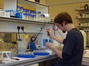
At the bench.

At the bench.

One of the approaches we often use is to visualize specific regions of the genome within living cells. First, a specific DNA binding protein ( called “LacI”) is genetically fused to GFP. Second, the DNA sequence recognized by LacI (called “lacO”) can be genetically integrated into any location of the genome. Since I study DNA replication, I am particularly interested in the site of the bacterial chromosome where DNA synthesis is initiated (called the “replication origin”). Third, fluorescent dyes are added to cells that bind to the cell membrane and the DNA. Finally, we utilize our fluorescent microscopes to visualize the location of replication origins within individual cells. In the image shown, the live bacterial cells contain chromosomes that are in the process of being replicated, and therefore they have duplicated and separated their replication origins!! This image also emphasizes the fact that although bacteria lack the organelles found in eukaryotic cells, they are nonetheless highly organized (notice how the replication origins are characteristically located at the outer edge of each chromosome).

The GFP protein from jellyfish can be used to fluorescently tag proteins in vivo. Fluorescence microscopy can then be used to localise the tagged protein within the bacterial cell.

The GFP protein from jellyfish can be used to fluorescently tag proteins in vivo. Fluorescence microscopy can then be used to localise the tagged protein within the bacterial cell.

Well that’s it for my first ICaMB blog! I hope you enjoyed hearing about how I became interested in bacterial genetics and about my work on bacterial DNA replication. Please feel welcome to contact me if you have any questions or if you would like further information regarding my research.


Links

Royal Society URF http://royalsociety.org/grants/schemes/university-research/

Centre for Bacterial Cell Biology (CBCB) http://www.ncl.ac.uk/cbcb/


A ‘tail’ to tell – Lakey lab discovery could lead to a new class of antibiotics


Dr Chris Johnson

“Most of us know that we should wash our hands after being around animals but do most of us know the reasons why? As a researcher who spends most of his time in the lab killing E. coli, using E. coli specific antibiotics I should be well aware of the dangers of this often underestimated Gram-negative bacteria. However when my youngest daughter contracted E. coli O157 after visiting an agricultural show in Scotland in 2011 (even though we had followed the hand washing procedure), I realised that I did not appreciate how nasty this bacteria can be. Thankfully after 6 weeks in hospital including a lengthy stretch on dialysis my daughter made a full recovery but not everyone is so lucky.”

Professor Jeremy Lakey

The urgent need to develop new drugs to target pathogenic bacteria has been a theme of the ICaMB blog since its inception in early 2013.  However, these words from Dr Chris Johnson, a postdoc in Jeremy Lakey’s laboratory, bring home the seriousness of the problem.  Fortunately, Chris is in a position to do something about this.

Many of you will have realised that last week saw another potential breakthrough in ICaMB’s ongoing ‘War on bacteria’ (and here, here and here).   This time the PI making the news, with data demonstrating the possibility of a whole new class of antibiotics, was Professor Jeremy Lakey.  And when we say making the news, we mean that literally. The ITV interview of Jeremy and his team can be viewed here

This story has made news around the world and has been featured in newspapers in Australia and India (and here), as well as closer to home.  Here is the official university press release.

So what’s going on?  If you want to read the primary paper it is here

The unstructured domain of colicin N kills Escherichia coli. Mol Micro 89:84-95

Chris Johnson, who was the lead author on this manuscript explains:

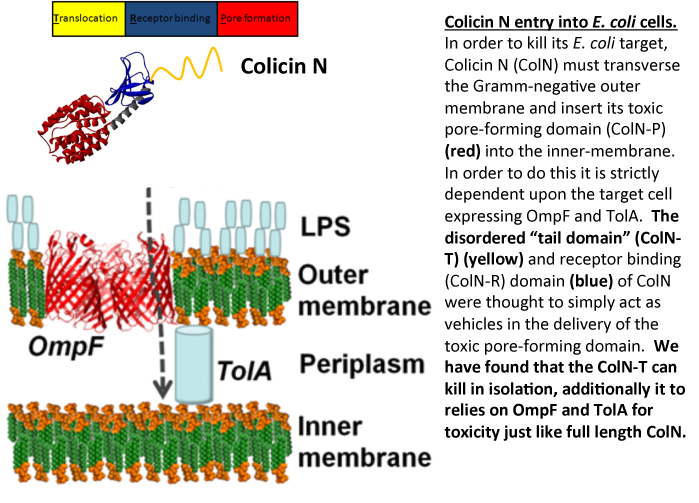
E. coli produces protein antibiotics called colicins which are used to kill E. coli and closely  related bacteria in the eternal bacterial arms race. In order to further understand how one of these, colicin N (ColN), works, we dissected the protein into its individual domains to see how each part behaved in isolation. Quite by chance we found that part of the protein, THE TAIL, was actually toxic to them. Although far less efficient than the entire ColN molecule, it remains specific for E. coli and furthermore, the specificity is housed within an intrinsically unfolded domain (a domain which has no defined 3D structure). Although this is a very basic discovery in its early stages, it allows us to appreciate novel mechanisms to kill E. coli.” (see the full version of this at the bottom of the page)

Some more about Jeremy

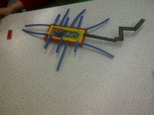
Any of you who were bioscience undergraduates in Newcastle will know Jeremy Lakey from his famous recreations of protein structure using party balloons.  Others may know him for co-founding Orla Protein technologies.  Some of us know him as a man who will always buy his round in the pub.  All of us know him as a great scientist and colleague.  Furthermore, Jeremy is a leading supporter of Leading Edge and recently a group of 6 Year 9 school pupils from St Cuthberts RC School looked at how ColN acts against E.coli when you start changing the amount of salt they are grown in. As many ICaMB scientists may know Jeremy also runs a workshop with Ponteland Community High School to explore bacterial shape and their surfaces using LEGO.

But did any of us think that one day Jeremy Lakey may SAVE THE WORLD from antibiotic resistant bacteria? Possibly.

The detailed science

Most Gram- negative bacteria produce protein antibiotics which are used as weapons in the battle between competing populations of bacteria.  E. coli produces protein antibiotics called colicins which are use to kill E. coli and closely related bacteria.  Once colicins are released into the extracellular milleu they dock onto their targets via specific outer-membrane receptors and then seek out an internal, periplasmic, binding partner (the Tol or Ton proteins) which helps them translocate into the cell.  We study colicin N (ColN) which comprises of an intrinsically unfolded N-terminal translocation (T) domain, involved in TolA and OmpF binding.  Its central receptor binding (R) domain binds lipopolysaccharide whilst its C-terminal 200 amino acids define the cytotoxic pore-forming (P) domain. This latter feature is common to all pore-forming colicins and forms a channel in the inner-membrane causing K+ release and cell death.  Other colicins have C-terminal domains which display cytotoxic activities that include DNAase or RNase activity.  Irrespective of the particular cytotoxic activity, all colicins are comprised of three domains (T-R-P) and it was assumed that the sole role of the T and R domains was to deliver the cytotoxic C-terminal domain across the outer-membrane.

In order to investigate the mechanism of ColN activity we dissected the protein into its individual domains.  We were attempting to block the toxic activity of full length ColN by pre- incubating E. coli cells with the intrinsically unfolded T-domain.  The rationale behind the experiment was that we could block all the available receptor sites on the E. coli target cells by saturating with T-domain, such that when the full length ColN was added to cells it would be non-toxic, as all the essential receptor binding sites would be already sequestered.  However rather than protecting the cells, T-domain was found to be toxic and like full length ColN provoked K+ efflux.   Although less efficient than full length ColN, T-domain is strictly dependent upon the same receptor proteins, OmpF and TolA for killing.  Since these receptors are only found in E. coli-like bacteria, T-domain displays the unusual combination of a generic killing mechanism coupled with extreme specificity housed within an intrinsically unfolded domain.

Links

Jeremy Lakey’s University home pagehttp://www.ncl.ac.uk/camb/staff/profile/jeremy.lakey

Follow Jeremy Lakey on Twitterhttps://twitter.com/JeremyLakey

ICaMBhttp://www.ncl.ac.uk/camb/

The Centre for Bacterial cell Biologyhttp://www.ncl.ac.uk/cbcb/

The official Newcastle University press release: http://www.ncl.ac.uk/press.office/press.release/item/chance-finding-could-lead-to-new-antibiotics

Link to ITV story:

http://www.itv.com/news/tyne-tees/story/2013-07-05/breakthrough-in-combatting-bacterial-infection/

The ‘Australian’ storyhttp://www.theaustralian.com.au/news/breaking-news/tail-could-be-used-for-new-drugs/story-fn3dxix6-1226674131684

The ‘Times of India’ Storyhttp://articles.timesofindia.indiatimes.com/2013-07-06/science/40406695_1_escherichia-coli-e-coli-protein

The Northern Echo storyhttp://www.thenorthernecho.co.uk/news/10524003.Scientists__chance_find_may_develop_new_generation_of_antibiotics/

Jeremy’s company, Orla Proteinshttp://www.orlaproteins.com/about-orla/the-board.aspx


Exploding bacteria for science!!!

As the Chief Medical Officer, Professor Dame Sally Davies, highlights today “danger posed by growing resistance to antibiotics should be ranked along with terrorism on a list of threats to the nation”. Professor Dame Sally Davies said diseases are evolving faster than the drugs that exist to treat them and antibiotic resistance is “a ticking time bomb“.

This is a subject of great interest to scientists in ICaMB, particularly the Centre for Bacterial Cell Biology, which brings together a world-class group of scientists researching bacterial physiology and the host response to bacterial infections. A major focus of this research involves:

  • Exploring alternative targets for antibiotic development
  • Understanding how antibiotics attack bacterial cells
  • Investigating how bacteria overcome such an attack

Tonight (March 11th 2013), work from the group of Kenn Gerdes and Etienne Maisonneuve, a post-doc in his group, will be featured on the BBC programme “Bang Goes the Theory” in an episode about Antibiotics.

Kenn and Etienne’s research focuses on persister cells, bacterial cells that can tolerate and survive attack by antibiotics.  Importantly, ALL bacteria analysed so far generate “persister cells” and understanding this is key to understanding how bacteria avoid antibiotic attack. “Bang Goes the Theory” will show a movie showing how these persister cells are identified in a bacterial population.

Penicillin inhibits synthesis of the bacterial cell wall, causing the cell to explode (or ‘lyse’) due to the high pressure inside the cell.  This is why penicillin and similar drugs are very effective in curing infections caused by penicillin-sensitive bacteria. In the movie, see how the cells suddenly explode when penicillin is added but notice how one cell, the persister cells (darker cells not exploding on the left panel) are surviving.

These persister cells evade killing by antibiotics because they grow extremely slowly. Persisters are proposed to be one explanation for infection relapses or chronic infections so Kenn and Ethienne’s work is extremely important for understanding how we should use antibiotics.

Microfluidic chamber used to make the movie

To do this work, Etienne used state-of-the-art technology – microfluidics – to follow the growth of individual bacterial cells under a microscope. These devices are smaller than a penny coin and the chambers where the bacteria are grown can be less than 1 mm across. This technique allows us to grow bacteria in one condition but, at a flip of a switch, change it and watch the response, as seen in the movie.

Year 9 student working on one of CBCB's microscopes


CBCB academics have used the ability to explode Escherichia coli to explore the what, when and how of antibiotics with Year 9 school students as part of the University engagement program Leading Edge.

With these students, we have developed a protocol to allow them to observe E. coli in the act of exploding after adding penicillin.

Exploding E. coli. Taken by Seaton Burn Community College Year 9 students

Persistence is not Resistance: It is important to understand the difference between these two terms. Antibiotic resistant and sensitive bacteria are able to generate persister cells, that are not effected by antibiotic attack. Antibiotic Resistance is a trait acquired by the whole population.

The Scientific Specifics: Over the last few years, several scientific breakthroughs made by the Gerdes group have, for the first time, given insight into how bacteria control the switch to slow growth and persistence. Persister cells can survive penicillin because the bacteria hibernate for a period, during which they don’t synthesize their cell wall.  They can then “wake up” when the antibiotic treatment is over, causing a new infection. In young and healthy people this is usually not a problem, because the rare non-growing bacteria are removed by the immune system. However, elderly individuals or those with a weakened immune system, it is often not efficient enough to permit clearance of the rare bacteria that survive the treatment, allowing the infection to “break out”.

The Gerdes group has shown that a certain class of gene that inhibits cell growth are turned on in one cell per 10,000. These discoveries open avenues to generate novel antibiotics and treatment regimes in the future. However, before that, their group is investigating if similar mechanisms allow pathogenic bacteria, such as Mycobacterium tuberculosis, to evade killing by antibiotics.


Institute of Cell and Molecular Biosciences
The Centre For Bacterial Cell Biology
Professor Kenn Gerdes
Bang Goes the Theory
Leading Edge

Bulging bacteria and the origins of life

Jeff (left), Romain (centre) and Yoshikazu (right), the team of researchers behind these exciting discoveries


In a paper published this week in CellJeff Errington’s team in ICaMB, have discovered new insights into the origin of life on Earth.


Jeff and his team share their results

Bacteria were the first organisms to appear on planet earth. Almost all modern bacteria have a tough protective shell called a cell wall. The structure of the wall and the mechanisms used by cells to manufacture it are conserved, suggesting that the wall was invented right at the beginning of bacterial evolution, and, therefore, when the first true cells emerged.

Production of cell wall is carefully regulated by complex machineries that allow the cell to enlarge and then divide in a controlled manner, all the time maintaining the integrity of the wall intact.

Despite its importance, it seems that many modern bacteria can survive cell wall loss under certain very special conditions, such as when they are treated with certain antibiotics that interfere with its production, like penicillin. Not only that, but a few years ago my lab showed that these “L-form” cells (named after the Lister Institute in London where they were first described) no longer need the complex mechanisms normally needed for bacterial growth and division. Instead, they grow by extrusion of irregular tubes or blebs of cytoplasm, that pinch off into daughter cells.

Our team – me, Yoshikazu KawaiRomain Mercier – has been working on this problem for some time. “Studying L-form biology is a real technical challenge, and this work could not have succeeded without the strong collaboration established between us“, says Romain. As Yoshikazu explains: “we developed a very simple genetic system to isolate mutations enabling L-form development from non-viable protoplasts.

We are excited because we think we have now solved the mystery of how L-forms grow and divide. Our latest results, published in Cell, show that the mechanism is remarkably simple: it requires only that cells make excess amounts of membrane – the thin porous layer that acts as the outer boundary of all cells, including our own.

Increasing the membrane surface area beyond the amount needed to contain the cytoplasm causes the cell to buckle and distort. Eventually, this leads to pinching off of membrane bags that are ill formed but nonetheless viable “baby” cells.

Time-lapse photography representing the division of B. subtilis without cell wall (L-form). The images were obtained using light microscopy. Scale bar: 3 μm

At first, we thought this mechanism was too simple to be true, we changed our minds when we were alerted to amazing experiments being done by several groups working on the origins of life, particularly Jack Szostak at Harvard, Saša Svetina in Ljubljana and Peter Walde in Zurich. These groups have been wondering how primitive cells could have arranged to grow and divide efficiently without spilling all of their contents. They recently found that simple membrane bags, called “vesicles”, can be induced to grow and reproduce into multiple smaller vesicles, in the test tube, just by increasing their surface area.

So, in explaining how the bizarre L-form bacteria manage to survive the loss of their beloved cell wall, we think we may now also have glimpsed how the first primitive cells could have duplicated themselves at the dawn of life on earth.

Jeff Errington 
Director of the Centre for Bacterial Cell Biology


Cell paper: http://www.cell.com/abstract/S0092-8674(13)00135-9
Cell website: http://www.cell.com/home see PaperFlick
Newcastle University Press Release:http://www.ncl.ac.uk/press.office/press.release/item/how-did-early-primordial-cells-evolve#.US-chen77jQ

Soapbox Science guest blogpost: http://www.blogs.nature.com/soapboxscience/2013/02/28/social-media-from-an-institutional-perspective-why-are-we-on-there

ICaMB website: http://www.ncl.ac.uk/camb/
Facebook: https://www.facebook.com/pages/ICaMB-Newcastle/416200498466481
Twitter: https://twitter.com/ICaMB_NCL
YouTube: http://www.youtube.com/channel/UCSuZgA6URiXTUoHq1tMe-PQ