Probity in Online Exams – Success at TEPARG for SDS and FMS TEL

This post highlights the work done by SDS and FMS TEL to support remote exams over the past few years. Successful techniques for keeping students informed and supported are discussed, including student voice.

Work done by the School of Dentistry and FMS TEL Team won first prize at the Trans-European Pedagogic Anatomical Research Group Hybrid Conference this year, for the presentation on ‘Adopting a flexible approach to professional anatomy spotter exams during COVID’. You can read the internal news item on Sharepoint.

This work was also subsequently presented at the Newcastle University Learning and Teaching Conference 2022. Newcastle Staff can view the poster here for an overview.

This work centres on how exams subject to oversight from professional bodies – in this case the General Dental Council – could be run with adequate probity when these could not be undertaken in person.

The first element of this is to run the exams ‘live’ rather than as a 24h format. This meant that teaching staff and professional staff could be on hand to resolve any technical difficulties or make invigilation decisions.

A variety of question designs were also thought through. The final format was a ‘stimulus’ question type on Canvas, allowing an image to be shown with answer options alongside. Now this question type can also be used with Inspera exams.

The support of students with SSPs was also a key consideration, and rest breaks were granted across the board when exams were very lengthy. Students were asked what they thought about the probity measures put in place, such as the use of an exam declaration, and checking responses which may have been copy-pasted. Overall, the response was positive.

A key element in the smooth running of these exams was the preparation offered to students beforehand, such as clear messaging via email and the opportunity to practice with the exam environment before undertaking their summative assessments. 99% of students agreed that they had been well-supported throughout the process and during the exams themselves via the Zoom exam hotline. Most calls were students double-checking their responses had been submitted, rather than having technical issues.

Learning from these exams has already been carried forward for digital exams running through the new Inspera exam system, and the confidence staff and students have in the procedure means that any future changes to circumstances will be much easier to navigate for these teams and cohorts.

Exploring 3D Anatomy – The University of Cape Town Launch

This week students from the University of Cape Town have joined the pilot of the Exploring 3D Anatomy course.

The second pilot of the Exploring 3D Anatomy course is now live, with students participating at the University of Cape Town in South Africa.

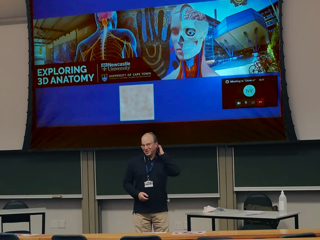
Leonard Shapiro presented an introduction to the course on 18th March to a class of second-year medical students – many of whom joined the class remotely via Teams.

Photo of a presentation Exploring 2D Anatomy.
Leonard presents the course
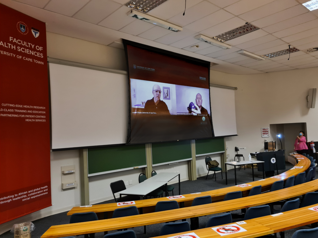
Photo of a classroom with Zoom call showing on a projection screen.
An example of one of the course exercises is shared
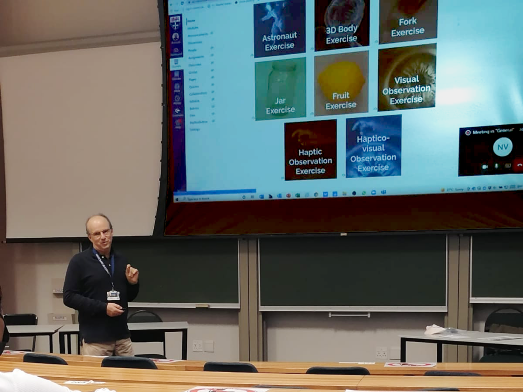
Photo of presenter and project screen showing exercises.
Leonard presents the course exercises
Dr Iain Keenan in the course trailer

The course trailer was also shown, and students were invited to sign up using a QR code posted on the slide show as well as on posters in the lecture theatre. Senior lecturer in Anatomy, Dr Geney Gunston, also posted the sign-up link for the students on VULA, the university messaging system.

Many students have signed up, and we look forward to working with them over the next few weeks as they Explore 3D Anatomy from home.

After this pilot and a further development phase, we hope to make the course more widely available to many more students.

Images provided by UCT medical student Mulaudzi God-mother Matodzi-muswa and Leonard Shapiro.

First Steps into 3D scanning

This year, 2021/22, FMS TEL took their first steps into 3D scanning to find a solution for a project in Dentistry.

Below you will find a summary of our progress to date, with further developments to come as we learn the craft of creating 3D digital images.

Equipment

  • Artec Leo scanner
  • Artec Studio 15 editing software
  • Sketchfab hosting

Artec Leo Scanner

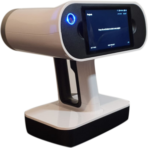
This is the Artec Leo 3D image scanner. It is a structured light scanner, using light to create geometrical shapes from objects to produce digital 3D versions of real world objects.

The Artec Leo is wireless and has an inbuild screen which you can view the progress of your scan on, rather than having to view on a computer screen like the other Artec scanners. You can store scans onto a micro SD card up to 256gb.

Artec Leo front
Artec Leo back

Find out more about he Artec Leo scanner: https://www.central-scanning.co.uk/product/artec-leo/

Artec Studio Editing Software

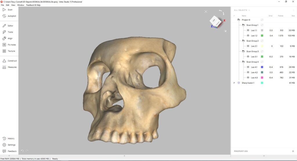
Artec Studio is used to edit scans, taking you through various processes to create a scan that is ready for online viewing or 3D printing.

Below is a screenshot from Artec Studio 15


Sketchfab Hosting

Sketchfab is one of a number of sites where you can host 3D scans.

Below are some examples of scans we have created and uploaded to Sketchfab

https://skfb.ly/osuCn

https://skfb.ly/osvxV

Exploring 3D Anatomy – A New MOOC Developed with FMS TEL

Over the past few months, the FMS TEL team have been working on bringing a Massive Open Online Course (MOOC) to life. The course, Exploring 3D Anatomy, is an active, hands-on, and engaging online course now available to Newcastle students and staff! The course was designed by Dr Iain Keenan of Newcastle University and Mr Leonard Shapiro of the University of Cape Town.

3D spatial awareness is a cognitive function. Improving it improves students’ 3D visualisation ability and spatial skills in anatomy learning.

In our experience, medical, dental, and other healthcare students can experience significant challenges in 3D spatial anatomy. Because of the three-dimensional arrangement of the human body, student spatial awareness can be a major influence on their anatomical education. In this online course, students can practice several  easy-to-follow, hands-on exercises that we have designed to address and improve 3D spatial awareness. Video demonstrations by Iain and Leonard guide students through each activity, which involve the use of  readily available household objects such as a piece of fruit, a jar, or a fork.

As simple as these exercises are to follow and carry out, the effect of such activities on improving 3D spatial awareness can be notable. What’s more, the exercises can be enjoyable too!

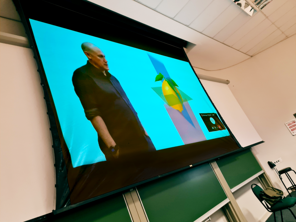
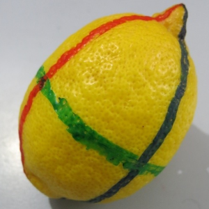
The Fruit Exercise: a guided dissection of this 3D object can improve 3D spatial understanding of the anatomical planes.

The practical exercises in the course are demonstrated by Iain and Leonard on video, allowing students to access the content at their own pace. These videos show the exercises in detail and allow students to hear the conversation as the exercise unfolds. Videos are short and simple to follow, and have been captioned by the team to ensure clarity.

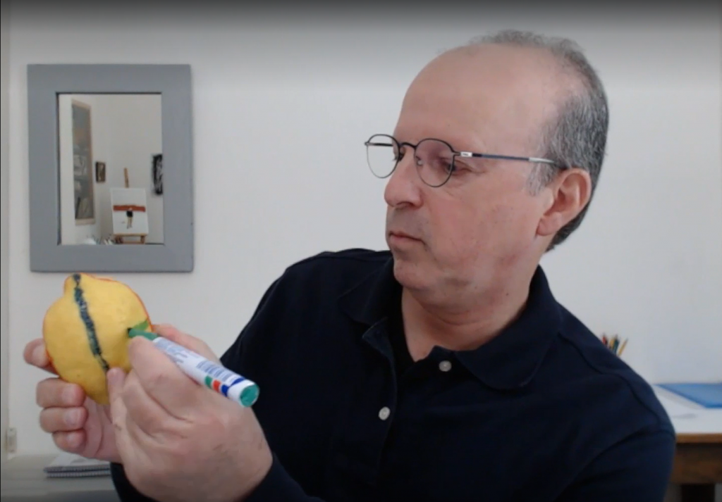
Leonard Demonstrating the Fruit Exercise with a Lemon
Iain Starting the Fruit Exercise with a Lime

All Newcastle University staff and students can join the Canvas course, which is structured in three parts and requires around 4 hours of activity in total. We hope to expand access to an extended version of the course in 2022/23. For further information, please contact Dr Iain Keenan.

Contacts 

Dr Iain Keenan, Senior Lecturer in Anatomy, School of Medical Education, Newcastle University 

Mr Leonard Shapiro, Observation and Spatial Awareness Teacher, Department of Human Biology, University of Cape Town 

Further Reading 

This course is supported by the following research: 

Backhouse, M., Fitzpatrick, M., Hutchinson, J., Thandi, C.S. and Keenan, I.D. (2017), Improvements in anatomy knowledge when utilizing a novel cyclical “Observe-Reflect-Draw-Edit-Repeat” learning process. Anat Sci Educ, 10: 7-22. https://doi.org/10.1002/ase.1616

Ben Awadh, A., Clark, J., Clowry, G. and Keenan, I.D. (2021), Multimodal Three-Dimensional Visualization Enhances Novice Learner Interpretation of Basic Cross-Sectional Anatomy. Anat Sci Educ. https://doi.org/10.1002/ase.2045

Branson TM, Shapiro L, Venter RG. Observation of Patients’ 3D Printed Anatomical Features and 3D Visualisation Technologies Improve Spatial Awareness for Surgical Planning and in-Theatre Performance. Adv Exp Med Biol. 2021;1334:23-37. Available at https://pubmed.ncbi.nlm.nih.gov/34476743/

Reid, S., Shapiro, L. and Louw, G. (2019), How Haptics and Drawing Enhance the Learning of Anatomy. Anat Sci Educ, 12: 164-172. https://doi.org/10.1002/ase.1807

Shapiro, L., Bell, K., Dhas, K., Branson, T., Louw, G. and Keenan, I.D. (2020), Focused Multisensory Anatomy Observation and Drawing for Enhancing Social Learning and Three-Dimensional Spatial Understanding. Anat Sci Educ, 13: 488-503. https://doi.org/10.1002/ase.1929

Digital Code for Teaching Anatomy Online – Joanna Matthan

Jo Matthan (Director of Academic Studies, School of Dental Sciences) talks about the teaching of Head and Neck Anatomy (DEN1101) within the School of Dental Sciences (SDS) at the Faculty of Medical Sciences (FMS), and how the move to online teaching necessitated the development of a specialised Digital Code around the use of cadaveric imagery.

Background

In Present-in-Person (PiP) teaching, students attending this foundational head and neck anatomy course would typically have access to the Dissecting Room for their learning. This would be delivered over a six-month period in their first year on either the Dental Surgery (BDS) or the BSc Oral and Dental Health Sciences degrees. This face-to-face time has been reduced to 12 hours from the approximately 50 hours of hands-on anatomy teaching delivered, which left a considerable amount of content to be covered in the digital format. In normal times, a code of conduct is signed by every student the first time they enter the restricted Anatomy and Clinical Skills Centre teaching facility. This was utilised in a digital format during the pandemic but, due to the major overhaul in teaching delivery, there was no way of reinforcing the messages contained within the code of conduct on a regular basis. It felt like a tall order to expect students to remember and understand a list of statements that is seemingly far-removed from them at the start of their anatomy learning journey, and to retain this volume of information in the digital era.

Due to the highly sensitive nature of working with donated cadaveric material and the associated professional standards and ethical considerations linked to this usage, it was necessary to develop a set of guidance to protect the dignity of donors, whilst simultaneously guiding educators, students and institutions on the manifold issues to consider when transitioning to online cadaveric teaching. The sensitive material (i.e., cadaveric images) would not normally be available to students on an ad hoc basis on their own devices. As this content still needed to be covered to ensure the healthcare professionals received all of the necessary training they needed to practice their profession safely and keep patients safe, questions arose around the potential for covert screen-capture, unauthorised viewing and wider sharing of cadaveric content. Such breaches of professionalism have widespread implications, not merely for the course and programme but potentially for the institution. It became clear that it was necessary to collate clear guidance for staff and students to steer through the digital landscape.

Developing an In-house Digital Code

An in-house digital code was developed as part of the SDS Digital Delivery Working Group – a student-staff collaboration that convened over summer 2020 to specifically mitigate for any anticipated issues that could arise from the shift to online delivery for both students and educators within the School. Three different digital codes were created, each in the form of a holding slide that could be utilised in (1) Cadaveric Anatomy Teaching, (2) Clinical Teaching and (3) Seminar or Lecture-based teaching situations. These slides were circulated to the FMS TEL Group and then circulated for feedback from the other Schools that grapple with similar concerns around donor dignity and patient confidentiality. A basic confidentiality agreement was later put in place institutionally, but this was not specific to anatomy or teaching involving potential breaches of patient information. It was felt that, in the new era of digital delivery, it would be more beneficial to reinforce the message of a common digital code at every learning encounter to clearly communicate to students the expectations and behaviours appropriate for this form of teaching. For this purpose, the Digital Code for anatomy was developed, with the aim of utilising it as a holding slide for every synchronous encounter, and as a recorded slide at the start of each non-synchronous session that included cadaveric content.

Digital Code in Practice

The Digital Code slide is displayed at the beginning of every teaching session, whether synchronous or non-synchronous. In synchronous sessions, it is used as the ‘holding slide’ displayed as students enter the video call. When teaching begins, the slide is also reinforced verbally to signal the start of the learning and shared professionalism code of conduct. In practice, it may feel that the points around ethical standards and professionalism are somewhat overemphasised but, given how important they are, the Digital Code does bear repeating. In non-synchronous sessions, a pre-recorded initial slide is added to every lecture recording to reinforce the message not to view the recording in public and to adhere to the digital code. This is added to all recordings from all contributors.

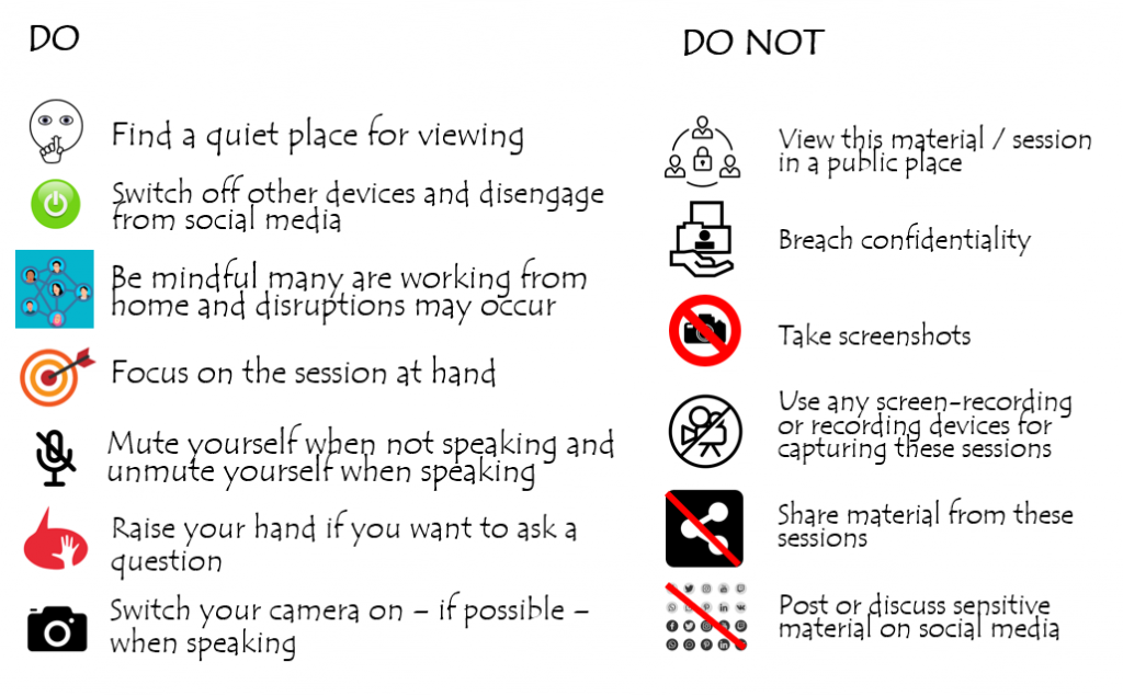
The slide shows 10 dos and don'ts. Do find a quiet place for viewing, switch off from other devices and social media, be mindful many are working from home and disruptions may occur, focus on the session at hand, mute yourself when not speaking and unmute yourself when speaking, raise your hand if you want to ask a question, switch your camera on if possible when speaking. DO NOT view this material/session in a public place, breach confidentiality, take screenshots, use and screen-recording or recording devices to capture these sessions, share material from the sessions, post or discuss sensitive material on social media
The Digital Code slide (download .ppt slide at the end of the post)

The Digital Code slide gives a simple overview of both the required professional behaviours for healthcare professionals and unacceptable behaviours, bringing the more abstract guidance document into clear actionable focus. As a result, students are very clearly aware of the professional expectations expected of them in their chosen field of study, and these regular reminders serve to reinforce this. This is much more effective than simply citing a document which may have only been seen once at the beginning of the course. The existence of this Code is beneficial not only for students who, it is hoped, develop a sense of responsibility with the access to sensitive content, but also for donors, teachers and institutions who can rely on clear guidance but also appreciate that institutions have pre-defined consequences for any breaches – of which none have been reported thus far. Students have displayed high levels of professional conduct throughout the anatomy teaching in SDS and have adopted the digital code without any apparent reluctance or challenges. It is highly likely that, once students return to physical classrooms, this practice will be continued and developed.

Taking it a step further: Developing National Guidance for Online Cadaveric Imaging

Due to manifold misunderstandings on the legislation and guidance around the use of cadaveric images, it became clear at an informal National Designated Individual (DI) / Head of Anatomy Forum (convened to improve communication during the pandemic and consisting of heads of anatomy units and DIs from across the UK, as well as representatives of the regulatory bodies from each country) that educators could benefit from a unified front with regards to digital cadaveric education. A small group from within this informal forum (consisting of representatives from Brighton Sussex Medical School, Newcastle University and University of Nottingham) collated anecdotal and professional experiences with patient confidentiality and social media guidance documentation and developed a three-pronged approach to using cadaveric content online. The first step was to search for guidance from the relevant professional bodies. The Human Tissue Authority (HTA), the national body who regulate teaching related to cadaveric specimens in England, is virtually silent on the use of images of a cadaveric nature, and decisions relating to how images can be used are made by the local HTA DI within institutions. There is also a paucity of guidance on image usage in this context from the devolved nations’ regulatory bodies/inspectors. Some institutions do have some guidance around social media and images, but there is no unified and unambiguous guidance on cadaveric teaching in the online era.

The draft guidance document was circulated to the DI Forum and to the HTA for comments and the final document amended with suggested changes. The current version, along with a suggested PowerPoint slide, is already utilised at SDS for teaching purposes. It has recently been presented at the March 2021 meeting of the Trans-European Pedagogic Anatomical Research Group (TEPARG), at which it proved very popular across the European countries represented, and has been widely lauded for its clarity and utility during this period of intense change. The guidance document, with the digital code appended to it, is now in use across many institutions nationally and internationally and is being reviewed for formal dissemination.

Resources

Human Tissue Authority

TEPARG – Trans-European Pedagogic Anatomical Research Group

Newcastle University Digital Etiquette Guide

Newcastle University Staff – Join our Canvas Community for access to all resources

Download the Slide and Guidance from our Canvas Community

3D Object Photography

3D object photography can have many purposes from selling products to interactive learning. It can be a complicated process, but we recently found a way to do it at home or in the office.

The thinking behind this for FMS is for the ability to show things such as models of parts of the body for example, which students away from the lab won’t have access to. They can interact with the object, and look 360 degrees around it. You can use a host such as Sirv (Links to an external site.) to create an interactive object.

Below is an example of what can be created. It can be viewed in full screen from the Sirv website:  https://tracyncl3d.sirv.com/Head/Head.spin (Links to an external site.) 

Would you like to try it yourself? You can find a guide on this in the FMS TEL Community

https://ncl.instructure.com/courses/30988/pages/3d-object-photography-at-home